
Page 25 - ISAKOS Newsletter 2016 Volume 1
P. 25
CURRENT CONCEPTS
Ulnar Neuritis
Another common elbow pathology in the throwing athlete is ulnar neuritis. The ulnar nerve arises from the medial cord of the brachial plexus and travels through several sites of potential compression. These sites include the arcade of Struthers, the medial intermuscular septum, the cubital tunnel, the two heads of the flexor carpi ulnaris, and in Guyon’s canal. In the throwing athlete, more common compression sites are the hypertrophied intermuscular septum, the medial head of the triceps, posteromedial osteophytes, and Osborne’s ligament.
For non-elite throwers, initial management of ulnar neuritis focuses on activity modification and rest. Nonoperative treatment is frequently unsuccessful in the elite throwing athlete due to the stresses across the elbow and nerve. Operative treatment for ulnar neuritis includes in situ decompression or anterior transposition. Throwing athletes are particularly predisposed to subluxation if the cubital tunnel is released without transposition. All sites of ulnar nerve compression should be released prior to transposition. Subcutaneous or submuscular transposition is then performed. Regardless of the transposition technique, the medial antebrachial cutaneous nerve, the ulnar vascular supply and the first motor bridge to the flexor carpi ulnaris must be preserved.
Flexor-Pronator Injury
The origins of the flexor-pronator mass are subject to overuse tendonitis since the flexor digitorum superficialis and flexor carpi ulnaris are prominent stabilizers during throwing. Flexor-pronator injury is generally treated conservatively with rest and non-steroidal anti-inflammatory drugs until symptoms resolve. The thrower is then rehabilitated with an emphasis on strengthening the flexor-pronator mass and graduating to an interval throwing program.
Medial Epicondyle Avulsion or Apophysitis
Medial epicondyle avulsion or apophysitis is more common in adolescent throwers and is low on the differential in the adult. Treatment is also conservative for medial epicondyle apophysitis, but if the medial epicondyle is completely avulsed, then open reduction and internal fixation is recommended. Athletes are permitted to heal through rest followed by a gradual return to throwing.
Treatment Strategies: Lateral Elbow Pain
Osteochondritis Dissecans of the Capitellum
Osteochondritis dissecans of the capitellum (OCD) remains an enigma. The spectrum of pathology can include injury to the subchondral bone and the development of frank loose bodies. Capitellar OCD is usually manifested with lateral elbow pain with both palpation and valgus stress.
Treatment for capitellar OCD is based on the severity of the lesion. Nonoperative treatment is usually commenced with an extended period of rest with no throwing. Nonoperative treatment is more likely to be successful in those with open physes rather than in mature individuals. Following resolution of pain, a strengthening and rehabilitative program is used to return the athlete to throwing.
Operative treatment of capitellar OCD is indicated in those who do not respond to nonoperative management and those with loose bodies. Debridement and removal of loose bodies can be performed with arthroscopy. Unstable lesions may be repaired; however, results are variable. In the event of an irreparable lesion, some authors have tried debridement with micro-fracture or osteochondral graft transplantation. The initiation of elbow strengthening and return to throwing is based on the procedures performed and the respective time to healing.
03
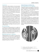
03 An incomplete olecranon stress fracture on presentation and at 3 months after nonoperative treatment.
02 Radiograph, T1 and T2 MRIs, and arthroscopic evaluation of osteochondritis dissecans of the capitellum.
ISAKOS NEWSLETTER 2016: Volume I 23
