
Page 24 - ISAKOS Newsletter 2016 Volume 1
P. 24
CURRENT CONCEPTS
Elbow Injuries in the Throwing Athlete: Location, Location, Location!
Imaging Studies
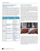
Standard radiographs of the elbow including anteroposterior, lateral and oblique views should be obtained. These radiographs can demonstrate stress fractures or avulsion injuries, osteophytes or presence of loose bodies. Stress fractures may not be apparent on routine radiographs and can be evaluated with CT or bone scans; however, MRI is the most commonly used advanced imaging modality because of its ability to detect stress injury in both bone and soft tissues. Ultrasound imaging is also gaining popularity for UCL evaluation due to its accessibility and affordability.
Table 1. Differential diagnosis based on symptom location.
Treatment Strategies: Medial Elbow
Ulnar Collateral Ligament Injuries
Treatment of UCL injuries depends on whether they are partial tears or complete ruptures. Partial tears are typically treated non-operatively with a period of rest for approximately three months with gradual return to play after therapy and a throwing program. Nonsteroidal inflammatory drugs are routinely used and some authors advocate for the injection of platelet rich plasma.
Operative indications for reconstruction of the UCL include complete ruptures of the ligament or in partial tears which remain symptomatic after nonoperative management. Primary repair of the ligament in adults was historically unsuccessful, however there may be a role in certain cases with early intervention. The original reconstructive technique described by Jobe et al utilized a free tendon graft through figure of eight bone tunnels in the medial epicondyle, detachment of the flexor pronator mass and ulnar nerve transposition. A later modification preserved the flexor- pronator mass and does not mandate transposition of the ulnar nerve.
02
An alternative method for UCL reconstruction includes the docking technique which splits the flexor carpi ulnaris and creates a single humeral tunnel. The graft is passed through the ulnar tunnels as in the Jobe technique and is then “docked” into the single humeral tunnel. The graft is tensioned with sutures tied over the humeral cortex. The docking technique was found to be biomechanically superior in the laboratory, but clinical outcomes have not identified superiority of either procedure. The largest UCL reconstruction cohort in the literature described 83% return to same level of competition.
Location
Tender to Palpation
Potential Pathology
Medial
Medial Epicondyle
Epicondylitis
Ulnar collateral ligament injury
Flexor-pronator mass injury
Cubital Tunnel
Ulnar Neuritis
Lateral
Radial Head
Valgus extension overload
Capitellar OCD
Posterolateral
Capitellar OCD Radiocapitellar plica
Posterior
Lateral olecranon
Stress fracture Osteophytes
Medial olecranon
Triceps avulsion Injury
Stress fracture Apophysitis Osteophytes
Posteromedial
Valgus extension overload
Osteophytes
22 ISAKOS NEWSLETTER 2016: Volume I
