
Page 23 - ISAKOS Newsletter 2016 Volume 1
P. 23
Elbow Injuries in the Throwing Athlete: Location, Location, Location!
Michael T. Freehill, MD
Department of Orthopaedic Surgery, Wake Forest School of Medicine Winston-Salem, NC, UNITED STATES
Austin V. Stone, MD, PhD
Department of Orthopaedic Surgery, Wake Forest School of Medicine Winston-Salem, NC, UNITED STATES
Elbow injuries in the throwing athlete can have a substantial impact on the player’s performance and may be highly detrimental to the player’s career. Unlike elbow injuries resulting from acute trauma, injuries in the thrower’s elbow tend to be chronic as a result of repetitive stress. The unique stress of throwing predispose the elbow to a subset of pathology reviewed in this article. In the throwing athlete, considerations for elbow pain include: ulnar collateral ligament injury, valgus extension overload, olecranon osteophytes, ulnar neuritis, olecranon stress fractures, flexor pronator injury, medial epicondyle avulsion fracture, capitellar osteochondritis dissecans, and loose bodies. These injuries are usually the cumulative result from repetitive stress during maximum internal rotation torque through the completion of the throwing motion with rapid elbow extension. This pattern was described by King and colleagues (1964) as the progression from medial tension and lateral compression overload to extension overload. The varying tensile, shear, and compressive forces can ultimately result in overuse injuries. Since many pathologies can be found in the throwing elbow, a thorough history of the timing and the location of the reported symptoms can help establish a differential diagnosis.
History and Physical Examination
The history should begin with an understanding of the throwing athletes sport, position, and level of activity or competition. The athlete’s pain should then be characterized based on its location, chronicity, associated neurologic or mechanical symptoms, and in the context of the athlete’s sport. The primary symptom may be decreased throwing speed, accuracy, or mechanical symptoms rather than pain. The timing and duration of the pain or symptoms should be characterized and potentially correlated with training regimen alterations, increased pitch counts or innings played, and any suspected traumatic event.
Physical examination in the throwing athlete should include an analysis of the general posture, resting position and muscle mass of the throwing and non-throwing arms. Elbow pain may be a manifestation of shoulder or scapular pathology, so both upper extremities should be examined in their entirety. The elbow carrying angle should also be evaluated. A normal carrying angle is 11° in men and 13° in women; although the carrying angle may be increased up to 15° in throwers. Asymmetric elbow range of motion in the throwing arm may be indicative of effusions, osteophytic change, or loose bodies.
The elbow should be palpated both before and during range of motion to assess for static and / or dynamic injury. The stability of the elbow can be assessed in varying degrees of flexion and extension and in pronation and supination. Since many of the injuries are related to chronic overload, a dynamic evaluation will best elucidate the pathology. Ulnar neuritis may be identified with a positive Tinel’s sign at the cubital tunnel. Integrity of the UCL can be tested with a modified milking maneuver and the moving valgus stress. The modified milking maneuver is performed with the shoulder in adduction and maximum external rotation. The examiner uses one hand to hold the elbow in 70° of flexion with the thumb on the medial joint line—70° of flexion was described in a cadaveric study to be the position of greatest valgus laxity in a sectioned UCL. The other hand is used to pull the thumb of the patient creating a valgus stress. Palpation of the joint line can determine joint space opening and presence of an end point. The moving valgus stress test described by O’Driscoll is performed with the shoulder at 90° of abduction and external rotation. The physician applies and maintains a constant valgus torque moving from a fully flexed elbow quickly into extension. A positive test results from apprehension, pain, or instability within the arc from 70 to 120 degrees. The elbow may also be evaluated with supination and rapid extension for assessment of posterior medial olecranon impingement.
01
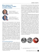
01 Loose body demonstrated on computed tomography scans and at the time of arthroscopic removal.
ISAKOS NEWSLETTER 2016: Volume I 21
CURRENT CONCEPTS
