
Page 16 - ISAKOS Newsletter 2016 Volume 1
P. 16
CURRENT CONCEPTS
Anatomic Deltoid Ligament Repair
Gian Luigi Canata, MD
Casale Centre of Sport Traumatology Koelliker Hospital
Torino, ITALY
Valentina Casale
Centre of Sport Traumatology Koelliker Hospital
Torino, ITALY
Introduction
The deltoid ligament, also known as the medial collateral ligament of the ankle, connects the medial malleolus to several tarsal bones.
Its superficial components include the tibiocalcaneal ligament, the tibionavicular ligament, the posterior superficial tibiotalar ligament and the tibiospring ligament.
Its deep layer is intraarticular, covered by synovium, and includes the anterior tibiotalar ligament (ATTL) and the posterior deep tibiotalar ligament (PDTL).
Both the superficial and the deep deltoid components work closely to stabilize the ankle against valgus and pronation forces. These components can be damaged after a rotational ankle fracture, ankle eversion injuries, or in the presence of chronic ankle instability, as well as in late stages of adult acquired flatfoot deformity. The most distinctive mechanisms of injury are pronation – abduction, pronation – external rotation and supination–external rotation of the foot.
Among the several classification systems developed over the years to stage the deltoid ligament injury, the Myerson’s classification is the most frequently used.
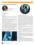
02
14 ISAKOS NEWSLETTER 2016: Volume I
01
Deltoid ligament injuries usually cause pain, tenderness and swelling on the medial side of the ankle. In the acute injury setting, it is important to thoroughly evaluate the syndesmosis for sprain or diastasis, and assess for fibula fractures. Injuries to the syndesmosis, which commonly affect the athletic population, are identified using a combination of palpation and stress tests. In the presence of an obvious syndesmosis injury (i.e., related to a fibular fracture), stress tests are not indicated and the patient typically ambulates with decreased power at push-off. Tenderness will be evident with palpation over the anterior- inferior tibiofibular ligament (AITFL) and pain will be perceived during external rotation and dorsiflexion of the ankle against the articular facet of the fibula.
If a fibular fracture is present, the medial side of the ankle must be assessed for the presence of a bimalleolar equivalent injury.
Pressure on the proximal portion of the fibula assesses the presence of a Maisonneuve fracture and hence the likelihood of syndesmotic and deltoid ligament disruption.
In the setting of chronic deltoid instability, the external rotation stress test also helps to evaluate the integrity of the deltoid ligament.
As the superficial component of the deltoid ligament crosses and stabilizes the subtalar joint, clinical evaluation of subtalar stability against valgus heel stress is also helpful. Be aware of global instability as well when examining the lateral ligaments. In severe chronic cases, valgus alignment and osteoarthritis can develop.
Radiographic Evaluation
Radiographic evaluation with standard plain radiographs may be useful for diagnosing a deltoid ligament injury due to a syndesmosis lesion, using the anteroposterior (AP), mortise and lateral views of the ankle. Weight bearing should be used if possible, though this can be difficult in the presence of fracture.
Clinical Presentation and Diagnosis
