
Page 14 - ISAKOS Newsletter 2016 Volume 1
P. 14
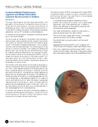
PEARLS & PITFALLS – SURGICAL TECHNIQUE
Combined Medial Patellofemoral Ligament and Medial Patellotibial Ligament Reconstruction in Children
Discussion
The main advantage of the technique presented is the possibility of carrying out the ligament reconstruction in skeletally immature patients and with the presence of predisposing factors. With the combined reconstruction, it is possible to stabilize the patella without performing other procedures such as ATT osteotomy and trochleoplasty.
In skeletally immature patients, procedures for preserving the physis are recommended.
For the fixation of the graft in the patella, tibia and femur, metal anchors were used due to their capacity to maintain the stability of the graft and eliminate the need for bone tunnels in the patella avoiding complications such as patella fracture and cartilage damage. The advantage of using anchors in the femur and tibia is the avoidance of drilling and bone tunnels near the physeal line, preventing injury. Kang et al demonstrated the effectiveness of using metal anchors for the reconstruction of the two bands of MPFL, with no evidence of failures or recurrence in the reconstruction. This technique allows for small and cosmetic incisions. After achieving skeletal maturity, patients should return for physical examination, as well as x-rays.
The fixation of the graft relative to the femoral distal physis remains controversial because of the publication of Shea et al in 2010, which demonstrated that the fixation point on the femur should be proximal to the physis. However, we fix the graft distal to the physis, inclined distally, slightly obliquely, to prevent the risk of perforation of the physis. We believe that the distal fixation to the physis is more reproducible with the use of fluoroscopy, as described by Kepler (2011), Ladd et al (2010) and Parikh et al (2013). When the fixation is positioned significantly distal to the physis, there is the possibility to overtension the patellofemoral joint in extension, which can be corrected by moving the point to an anterior position in the same proportion that it is placed distally.
In the tibia, the fixation of the graft at the proximal epiphysis instead of the tibial proximal metaphysis, avoids the risk of distal migration of the graft insertion with growth, which could cause a change in tension and function.
The reconstruction of MPFL combined with medial MPTL using flexor tendons as graft has shown satisfactory results. Summarizing the main Tips and Pitfalls in the combined reconstruction of MPFL and MPTL:
• It should be recommended to skeletally immature patients with anatomical risk factors such as generalized ligamentous laxity, knee hyperextension, instability in flexion and subluxation of the patella in extension with the contraction of the quadriceps.
• The single semitendinosus tendon may be used as a graft with preservation of its tibial insertion.
• The use of anchors avoids bone tunnels in the patella and near the physis of the femur and tibia.
• The identification of anatomic points with the aid of fluoroscopy.
• Appropriate tensioning of MPTL at 90° of flexion with similar tension to the patellar tendon identified by touch.
• And, lastly, avoiding the overtensioning of MPFL, which could cause the medial subluxation of the patella, patella baja and medial overpressure of the patellofemoral joint.
03
12 ISAKOS NEWSLETTER 2016: Volume I
