
Page 13 - ISAKOS Newsletter 2016 Volume 1
P. 13
Surgical Technique
Firstly, the arthroscopic procedure is carried out and the articular cartilage, the menisci and ligaments are inspected. The surgical procedure is performed with three small incisions. The first incision is made at the proximal medial tibia in order to identify the insertion of pes anserinus tendons. The semitendinous tendon is divided from the muscle-tendon junction, preserving the distal insertion site. The tendon must be at least 12-13 cm in length. The semitendinosus tendon was chosen for greater length and sufficient strength for the reconstruction. A second incision is performed at the medial border of the patella, including the landmark for the fixation of the graft. The last incision is made in the area between the adductor tubercle and the medial epicondyle with the aid of fluoroscopy for the identification of anatomical points of graft insertion.
After confirming the stabilization of the patella and full range of motion with complete flexion- extension, the graft is enhanced with the suture at the periosteum of the patella between the suture anchors to increase the area of patellar insertion, according to its anatomical characteristics (Fig. 2 and 3).
After surgery, the patient remains with a removable immobilizer for three weeks. Physical therapy is started on the second day after surgery with isometric exercises for the quadriceps. The exercises for range-of-motion and quadriceps strengthening are encouraged progressively and as tolerated after the 30th day after surgery. The load without crutches is allowed when the patient is able to walk without a limp. One month after surgery, knee flexion of more than 90° should be achieved. Sports activities are restricted for 4–6 months.
After preparing the graft with sutures at its free
end, a metal anchor is placed in the tibia with a
small incision in the region of the medial patellotibial
ligament insertion, proximal to the physis, between
the medial border of the patellar tendon and the
superficial medial collateral ligament, forming an
angle of approximately 20° with the patellar tendon,
proximal to the physeal line. The graft is fixed at this
point and then transferred percutaneously to the
distal incision from the medial border of the patella,
which is fixed with a metal anchor at 90° of knee
flexion with tension similar to the one perceived
in the patellar tendon, according to Hinckel at al.
A third anchor is positioned on the patella, at the
point between the middle and proximal thirds of the
patella in its medial border. Then the graft is passed
to the femur through the second layer of the medial
retinaculum of the patella, and the fourth anchor is
placed in the distal region to the physis, by means of
fluoroscopy, according to the study by Schöttle et al,
in the region between the medial epicondyle and the 02 adductor tubercle, between 5mm and 6.4mm distal
PEARLS & PITFALLS – SURGICAL TECHNIQUE
to the physis. Prior to the insertion of the anchor, the positioning must be checked in both planes, AP and true lateral. The graft is then secured with the knee between 45° and 60° flexion without excessive tension, reconstructing the medial patellofemoral ligament. The patella is tested during flexion- extension by observing its medium-lateral course, which should allow mobility between 25% and 50% of the patella without lateral displacement.
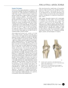
01 Insertion and reconstruction of medial patelofemoral ligament with quadricipital tendon and of medial patellotibial ligament with patellar tendon.
02 Reconstruction of the Medial Patellofemoral Ligament (MPFL) and the Medial Patellotibial Ligament (MPTL) with the semitendinosus tendon.
ISAKOS NEWSLETTER 2016: Volume I 11
