
Page 12 - ISAKOS Newsletter 2016 Volume 1
P. 12
PEARLS & PITFALLS – SURGICAL TECHNIQUE
Combined Medial Patellofemoral Ligament and Medial Patellotibial Ligament Reconstruction in Children
David Sadigursky, MD
Orthopedic and Traumatologic Clinic
COT Hospital Manoel Victorino-Salvador, Bahia, BRAZIL
Anatomical and biomechanical studies have shown that the Medial Patellofemoral Ligament (MPFL) is the main restrictor to the lateral translation of the patella. Numerous surgical techniques have been described in the literature for the reconstruction of the MPFL with favorable clinical outcomes. In addition to the MPFL, ligaments that contribute to the medial restriction of the patella are the Medial Patellotibial Ligament and the Medial Patellomeniscal one. The latter contribute to the restriction of the patella at angles above 30° of knee flexion. Philipot et at showed that the MPTL contribution increases in extension from 26% to 46% at 90° flexion. Besides contributing to patellar stability, the MPFL influences the tilt and the rotation of the patella. Hence, the reconstruction of the MPFL combined with the MPTL is important to maintain the normal kinematics of the patellofemoral joint throughout the range- of-motion. As found by the group of knee surgery of the University of São Paulo (IOT HC USP), the combined reconstruction with a secondary stabilizer (MPTL or MPML) could reduce stress on the rebuilt MPFL and thus increase the functional results and may be an attractive technique when the osteotomy of the Anterior Tibial Tuberosity (ATT) is not possible.
Various grafts have been demonstrated to be effective for the combined MPFL and MPTL reconstruction. Different ways of fixing these grafts have been described with favorable clinical outcomes, despite maintaining a rate of complications around 26.1% with both objective and subjective instability rates of 12%, according to the systematic review published by Shah et al in 2012.
In 1922, Galeazzi described a MPTL reconstruction technique in patients with open physis, in order to control the height of the patella in relation to the femoral condyle and transmit the contraction force from the quadriceps to the tibia. Giordano et al, in 2012, demonstrated the MPFL/MPTL combined reconstruction using the semitendinosus and gracilis tendons with a longitudinal bone tunnel on the patella and fixation on the femur with a metal anchor. The technique was indicated for skeletally immature patients with a high patella, trochlear dysplasia, increased TT-TG and ligamentous laxity.
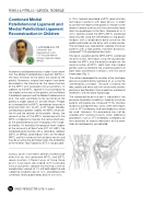
Hinckel et al published the MPFL/MPTL combined reconstruction technique using the quadriceps tendon for MPFL and the patellar tendon for the reconstruction of MPTL, both from their medial portion and transferred to the anatomical points of both tibial and femoral insertions, with the aid of fluoroscopy (Fig. 1).
The authors developed the variation of the technique previously published by Giordano et al, using the semitendinosus tendon. The graft is fixed to the tibia, patella and femur with the use of metal anchors previously positioned in their respective anatomical landmarks, with the aid of fluoroscopy.
The combined reconstruction is indicated in the presence of patellar instability in skeletally immature patients with patella alta, increased TT-TG, trochlear dysplasia and ligamentous laxity, when techniques such as ATT osteotomy and trochleoplasty cannot be used. However, this procedure may not be needed after skeletal maturity when additional procedures such as ATT osteotomy, osteotomy for the correction of angular deviations of the lower limbs or trochleoplasty are indicated.
01
10 ISAKOS NEWSLETTER 2016: Volume I
