
Page 10 - ISAKOS Newsletter 2016 Volume 1
P. 10
PEARLS & PITFALLS – SURGICAL TECHNIQUE
Hip Preservation
Surgical Technique
If the patient is known to have dysplasia or does not have a pincer lesion which requires rim resection, they are good candidates for the ‘inside-out’ technique.
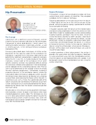
Following debridement of the labral base tear the labrum is lifted slightly off the rim at the site the anchor will be placed, and manual gentle rasping is performed to facilitate revascularization for healing.
Drill guide is placed under the labrum (at the chondro-labral junction) ensuring ample bone in which the anchor can take hold. Either straight or angled guides can be used according to portal trajectory and repair’s hour of the clock. Once the anchor (Nanotack, Stryker) has been secured, a labral base repair, or a repair with stitch around the labrum, can be performed. In order to complete a labral base repair, both suture limbs are retrieved through the labrum, in a vertical or horizontal mattress fashion, to the capsular side of the labrum using the arthroscopic suture passer (Nanopass, Stryker). Arthroscopic knot tying techniques are used to secure the labral repair.
In order to perform a labral advancement, the anchor is placed in the same fashion, but after the labrum is freed from its native attachment away from the chondrolabral junction. Once the labrum can be mobilized freely with a probe, it will be brought closer to the chondrolabral junction using the anchor suture limbs.
01a 01b
01c 01d
The Concept
Omer Mei-Dan, MD
Associate Professor
Hip Preservation Center
University of Colorado Denver/Boulder, CO UNITED STATES
Labral tears are a significant cause of hip pain, and are currently the most common indication for hip arthroscopy. Compared to labral debridement, labral repair has significantly better outcomes in both daily activities, as well as athletic pursuits, in the setting of femoral acetabular impingement (FAI).
Previously described repair techniques all utilize anchor placement on the capsular aspect of the acetabular rim and labrum, a position that is not always ideal and has the potential for significant complications, especially anteriorly where the rim is very thin. A shallow dysplastic hip, drilling trajectory, narrow width of the acetabular rim or some specific anatomic variations may generate difficulty during this commonly utilized anchor placement. Anchor breakage, anchor slippage into the surrounding (capsular side, IP bursa) soft tissue or penetration of the cartilage surface are among the most common complications. Furthermore, anatomical anomalies do exist in the acetabular labrum, where the native labrum does not create an adequate suction seal with the femoral head, reducing even distribution of cushioning synovial fluid, and increasing the forces within the joint. In anatomical cases such as these, the superior deviation of the labrum would only be worsened by the direction of pull from sutures coming out of an anchor placed behind the labrum, displacing the labrum from the correct anatomical position that much further.
We describe an intra-articular anchor placement technique, which provides a means to repair a torn labrum when more standard approaches are less ideal. This technique can also assist in labral advancement (puling the labrum towards the femoral head to create a more functional seal), when the native labrum fails to create a proper seal due to its location away from the femoral head.
8 ISAKOS NEWSLETTER 2016: Volume I
