
Page 17 - ISAKOS Newsletter 2016 Volume 1
P. 17
CURRENT CONCEPTS
The radiographic parameters of a syndesmotic injury are:
• Increased tibiofibular clear space: The distance between the medial border of the fibula and the lateral border of the posterior tibia as it extends into the incisura fibularis (normal value <6mm in AP and mortise views)
• Decreased tibiofibular overlap (normal values >6mm or >42% of the width of the fibula on the AP view; >1mm on the mortise view)
• Increased medial clear space: The distance between the lateral border of the medial malleolus and the medial border of the talus
03 04
Porter et al. (2014) stated that a syndesmosis injury may be present if there is more than 1 mm lateral subluxation or more than 5 mm supination between the distal fibula and tibia on the mortise view.
It is important to note that isolated deltoid rupture does not necessarily involve a widening of the medial clear space. If the syndesmosis remains intact, the lateral malleolus holds the talus in position. This type of injury can be difficult to assess with static radiographs, thus eversion-stress radiographs help to detect isolated deltoid injury, and latent syndesmotic injuries.
Finally, when a proximal fibula fracture is suspected (i.e., Maisonneuve injury), anteroposterior and lateral views radiographs of the entire fibula should be completed.
Computed tomography (CT) is more sensitive to detect minimal (2-3 mm) syndesmotic diastasis, not visible on plain radiographs.
Magnetic resonance imaging (MRI) is highly sensitive and specific for the diagnosis of syndesmotic injuries. It has been demonstrated that the widening of the medial tibiotalar clear space on the mortise view may not be closely associated with deep deltoid ligament injuries. For these reasons, MRI may be helpful for early and accurate diagnosis, mitigating the risk of chronic medial instability, ostheoarthritis and medial ankle impingement syndrome.
Indications for Surgery
The key to appropriate healing of the deltoid is stabilization of the syndesmosis.
When a syndesmotic injury occurs, treatment options depend on the severity of the lesion:
• Type 1: There is clinical evidence of a syndesmotic injury, without radiological signs. In this case, conservative treatment can be successful with weight-bearing allowed as tolerated.
• Type 2: There is radiological evidence of syndesmotic affection. A non- weight bearing cast is positioned for the first 30 days, then weight-bearing is gradually permitted.
• Type 3: An ankle syndesmotic diastasis occurs. In this case, surgical treatment is recommended to reduce and stabilize the syndesmosis.
Eversion ankle sprain and subsequent instability may cause partial or complete deltoid ligament tear, although surgery is rarely needed if the ankle mortise remains stable.
Syndesmotic injuries, even without fractures, involve the distal talofibular joint and can lead to ankle mortise instability. Complete deltoid ligament ruptures usually develop in combination with ankle fractures.
The possible associated fractures include supination-external rotation ankle fractures (Weber type B), pronation-external rotation ankle fractures (Weber type C) and fractures of the proximal fibula (Maisonneuve type). Since these injuries are typically associated with lateral ligamentous injuries, surgery is often recommended in case of fractures.
A deltoid ligament lesion or a combination of osseous and ligamentous lesions may develop in case of medial instability associated with a lateral malleolar fracture, caused by a medial malleolar fracture.
An entrapment of the deltoid within the medial gutter of the ankle may result from unstable bimalleolar ankle fractures, especially when the mortise remains wide medially after anatomic reduction and fixation of the lateral side of the ankle.
05
01 MRI: Deltoid ligament dislocated under the medial malleolus
02 MRI showing the fibula lying outside the syndesmotic lesion
03 Lateral incision for syndesmotic repair
04 Lateral side after reduction with screws
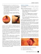
05 Medial side incision below the medial malleolus
ISAKOS NEWSLETTER 2016: Volume I 15
