
Page 18 - ISAKOS Newsletter 2016 Volume 1
P. 18
CURRENT CONCEPTS
Anatomic Deltoid Ligament Repair Surgical Technique
The patient lies in the supine position, the tourniquet is placed on the calf and inflated 100 mmHg above the systolic arterial pressure. In the absence of acute fractures, the stability of the syndesmosis is tested with external rotation and abduction stress under fluoroscopy. If a fibula fracture is present, this should be stabilized and repaired before stress testing of the syndesmosis and medial ankle.
The surgical approaches for the stabilization of the syndesmosis may be both medial and lateral.
The lateral approach is made to repair the fibula fracture and reduce and repair the syndesmosis, while the medial approach is used to repair a ruptured deltoid ligament. When the lateral approach is performed, use caution to avoid damage to the sural and superficial peroneal nerves. Carefully reduce any distal fibula fracture, reduce the diastasis and correct any malrotation. After the fibula reduction, the syndesmosis may be stabilized with two screws across four cortices. Then, it is possible to repair the AITFL, if completely torn.
When the medial approach is performed, a curvilinear skin incision distally and parallel to the medial malleolus is made. After the exposure and the incision of the most superficial layers, including the flexor retinaculum, the deltoid ligament is evaluated. If the deep ligament is compromised due to a syndesmotic injury, a primary repair of the ligament can be performed.
During this procedure, it is important to ensure that the posterior tibial tendon (PTT) and the spring ligament are intact and protected, otherwise the ankle joint stability will not be guaranteed.
The PTT sheath is incised and the tendon is retracted, in order to obtain a good inspection of the articular surfaces. Any possible cartilage damage must be debrided and drilled if necessary.
Using no. 1 Vicryl sutures, the deep fibers of the deltoid ligament are sutured and tied; then the superficial stitches are secured only after the osteosynthesis is completed and a definitive reduction and fixation on the lateral side is obtained, in order to avoid any possible future suture avulsion.
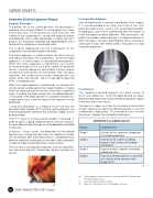
The final result, including joint reduction and new alignment, may be evaluated with fluoroscopy, before layered skin closure.
06
Postoperative Regimen
Joint immobilization is advised immediately after surgery. It is recommended even from the third to the sixth week postoperatively, using a non-weight bearing boot. Radiographic assessment is performed after four weeks to check the correct anatomic alignment. After two months, the patient may begin weight bearing in a hinged ankle brace. After nine weeks, running is permitted but always wearing an ankle tape. Finally, after twelve weeks, the patient can stop using joint protections.
07
Conclusions
The superficial deltoid ligament has been shown to resist talar abduction, while the deep deltoid has been demonstrated to be more closely related to rotational stability of the talus within the mortise.
The goal of surgery must be the anatomical restoration of both deep and superficial deltoid ligaments and the syndesmosis stabilization. This technique is less invasive than others, thereby it ensure optimal outcomes.
MYERSON’S CLASSIFICATION
STAGE
DESCRIPTION
Stage 1
Avulsion of the super cial component of the deltoid ligament
Stage 2
Rupture of the super cial deltoid ligament and avulsion of the deep component
Stage 3
Rupture of both super cial and deep deltoid ligaments
Stage 4
Rupture of deltoid ligament associated with rupture of the posterior tibial tendon (PTT)
06 Deltoid ligament detached from the medial malleolus. Medial repair with absorbable sutures.
07 Anatomic alignment after surgery
08 Myerson’s classification for deltoid ligament injuries
08
16 ISAKOS NEWSLETTER 2016: Volume I
