
Page 34 - ISAKOS 2020 Newsletter Volume 2
P. 34
CURRENT CONCEPTS
Posterolateral Corner Surgery: Tips & Tricks from Asian Surgeons
Femorofibular-Based PFL Anatomical PLC Reconstruction
Many knee experts have modified the anatomical reconstruction to improve and further replicate anatomy. There are still limitations on how well surgeons are able to recreate the native anatomical structures of the PLC. First, the PT normally provides both dynamic and static stability to the knee. In PLC reconstruction procedures, the popliteus is converted to a static stabilizer. The implications of this conversion are still to be investigated. Second, the native PFL that originates from the musculotendinous junction of the PT is hard to mimic. As a result, there is no clear consensus on the procedure to reconstruct the site of the PFL origin on the PT.
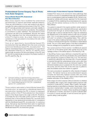
Yoon et al. described a femorofibular-based PFL reconstruction that was different from the more commonly performed tibiofibular-based PFL reconstruction3. The authors emphasized that it was important (1) to reconstruct all 3 structures of the PLC (i.e., the FCL, PT, and PFL), (2) to anatomically reconstruct the femoral insertion of the FCL and PT, and (3) to anatomically reconstruct the fibular insertion of the PFL and FCL (Fig. 4).
04 Schematic drawings of a right knee showing posteroanterior
(Fig. 4-A) and lateral (Fig. 4-B) views of graft fixation for anatomical PLC reconstruction with a femofibular-based PFL reconstruction. LCL = lateral collateral ligament (i.e., FCL), PFL = popliteofibular ligament, PLT = popliteal tendon.
Those authors advocated a femorofibular-based PFL reconstruction, as opposed to a tibiofibular-based PFL reconstruction, because the PFL is known to represent a static portion of the PT that extends from the posterior aspect of the fibular head to the origin of the PT. Therefore, to recreate the role of the static portion of the PT, that group believed that a femorofibular-based PFL reconstruction represents a more anatomical reconstruction in comparison with the tibiofibular-based PFL reconstruction. In a previous study, that group reported that there were no significant differences in clinical outcomes between femorofibular and tibiofibular-based PFL reconstruction groups.
Arthroscopic Posterolateral Capsular Stabilization
Isolated PLC injury is an uncommon knee pathology that could be a source of unexplained knee pain and dysfunction due to posterolateral rotational instability (PLRI). Ohnishi et al. devised an entirely arthroscopic approach for the treatment of isolated PLRI by stabilizing the posterolateral joint capsule along with the lateral meniscus on the rim of the lateral tibial plateau5.
The patient is placed in the supine position under spinal or general anesthesia, and a standard arthroscopic examination is performed through an anteromedial portal. The drive- through sign is used to evaluate the PLC injury by measuring the displacement of the lateral meniscus with use of a probe (Fig. 5-A). A mid-lateral portal is created posterior to the FCL and 1 cm anterior to the popliteal hiatus. Then, with the scope in the anteromedial portal being used to view the site, a shaver is introduced through the mid-lateral or anterolateral portal. The rim of the lateral gutter of the tibia is abraded to remove cartilage and is prepared for anchor placement.
Two suture anchors (Y-knot anchor; ConMed) are placed on the rim of the lateral gutter after partial removal of the cartilage surface of the lateral aspect of the tibia (Fig. 5-B).
A QuickPass Suture Lasso (Arthrex) is inserted through the mid-lateral portal, and the tip penetrates the posterolateral joint capsule from superior to inferior (Fig. 5-C). The wire loop is advanced sufficiently into the knee joint. A suture grasper is inserted through the anterolateral portal, and a wire loop and 1 strand of braided suture are retrieved together. The retrieved braided suture is passed into the wire loop outside the portal, and the other limb of the wire loop is pulled to pass the braided suture from inferior to superior.
The same procedure is repeated for other strands of braided suture. The lateral meniscus and posterolateral capsule complex are fixed with mattress sutures with use of the suture relay technique (Fig. 5-D), which is less invasive and enables restoration of the stability of PLC.
Knee range-of-motion exercise without restriction is initiated immediately after surgery. Non-weight-bearing exercise with crutches is continued for 2 weeks. After 4 weeks, full-weight- bearing exercise is permitted as tolerated. Open kinetic chain exercise is started after 5 weeks, and closed kinetic chain exercise is started at 10 weeks. Deep squatting and jogging are allowed at 3 months, and return to sports is allowed after 4 months if muscle strength is well recovered and sports- specific ability is well acquired.
32 ISAKOS NEWSLETTER 2020: VOLUME II
