
Page 32 - ISAKOS 2020 Newsletter Volume 2
P. 32
CURRENT CONCEPTS
Posterolateral Corner Surgery: Tips & Tricks from Asian Surgeons
Proper assessment and diagnosis of PLC injuries are essential for appropriate treatment. PLC repair and reconstruction are performed to restore knee stability. PLC reconstruction techniques can be broadly classified into non-anatomic and anatomic on the basis of the ligaments reconstructed and the position of the reconstruction tunnels. Anatomical reconstructions surgically reproduce the main 3 stabilizers to the PLC (the FCL, PT, and PFL) and their anatomical footprints. Non-anatomic reconstructions either do not reproduce all 3 structures or do so by means of non- anatomic attachments.
The anatomical PLC reconstruction technique proposed by LaPrade in 2004 has been adopted by surgeons worldwide, with modifications in fixation and graft choice. We present a collection of tips for assessing and treating PLC injuries as reported by Asian surgeons over the last 10 years1.
Lateral Gutter Drive-Through Sign and Femoral Popliteus Peel-Off Lesion
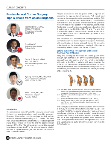
Feng and colleagues described the lateral gutter drive- through sign, defined as the arthroscopic finding of a lateral compartment joint opening of >1 cm, which is consistent with laxity of the FCL. In patients with a positive sign, the arthroscope passes into the posterolateral compartment through the interval and lateral femoral condyle at 30° of knee flexion, which indicates posterolateral rotatory instability of the knee (Fig. 1).
01A The lateral gutter drive-through test. The arthroscope is inserted through the interval between the popliteus tendon and the lateral condyle into the posterolateral compartment of the knee.
01B Type-I femoral peel-off lesion (isolated PT tear from the femoral insertion site).
01C Type-II femoral peel-off lesion (combined PT and FCL tears). 01D Type-III femoral peel-off lesion (complex tear with both the PT
and FCL torn from the femoral insertion site with complex mid- substance injuries).
The same group described femoral popliteus peel-off lesions in a study of patients with acute PLC injuries (Fig. 2)2. They reported that 82% of patients with peel-off lesions showed conclusive evidence of femoral insertion separation and discontinuity. Physical examination revealed external rotation of >10° in 84% of patients and positive varus instability in 73%. Intraoperatively, arthroscopic findings of acute avulsion and a positive lateral gutter drive-through sign were found in 94% of patients.
Introduction
Yee-Han Dave Lee, MD, MBBS, FRCS(Orth) Changi General Hospital SINGAPORE
Feng Hua, MD
Beijing Jishuitan Hospital Beijing, CHINA
Sachin R. Tapasvi, MBBS, MS, DNB, FRCS
The Orthopedic Specialty Clinic Pune, INDIA
Kyoung-Ho Yoon, MD, PhD, Prof.
Kyung Hee University Hospital Seoul, SOUTH KOREA
Soshi Uchida, MD, PhD
Wakamatsu Hospital Fukuoka, JAPAN
The posterolateral corner (PLC) of the knee was once an area that was poorly understood and much feared by surgeons. However, this area has been increasingly studied, and we now have a better understanding of PLC injuries. The three main structures of the PLC are the fibular collateral ligament (FCL), the popliteus tendon (PT), and the popliteofibular ligament (PFL). The FCL is the primary restraint to varus at lower flexion angles, the PT is the principal external rotation stabilizer at higher flexion angles, and the PFL stabilizes the knee in internal rotation.
30 ISAKOS NEWSLETTER 2020: VOLUME II
