
Page 33 - ISAKOS 2020 Newsletter Volume 2
P. 33
The lateral side of the knee is exposed, and anatomical landmarks are identified as described by LaPrade. The femoral sockets for the popliteus and FCL are drilled first, followed by the oblique fibular tunnel. The tibial tunnel for the popliteus is drilled for a length of 25 mm with a retro drill cutter. Graft passage is exactly reversed from the technique described by LaPrade.
The doubled 25-mm graft is initially passed in a blind tibial popliteus socket (Fig. 3). Tensioning is achieved by means of an adjustable loop with cortical tibial button fixation. Subsequently, the 2 free ends are used to reconstruct the 2 limbs of the PLC. The short free end of the graft is routed proximally to the popliteus femoral tunnel under the iliotibial band (ITB) and is fixed with a bioabsorbable screw with the knee in 90° of flexion and neutral rotation with a lateral closing force (Fig. 3).
CURRENT CONCEPTS
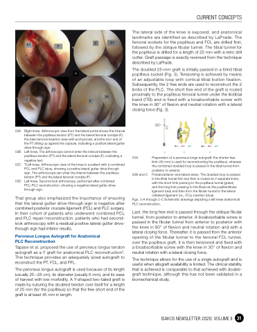
02A Right knee. Arthroscopic view from the lateral portal shows the interval between the popliteus tendon (PT) and the lateral femoral condyle (F), the bare femoral insertion area with ecchymosis, and the torn end of the PT sitting up against the capsule, indicating a positive lateral gutter drive-through sign.
02B Left knee. The arthroscope cannot enter the interval between the popliteus tendon (PT) and the lateral femoral condyle (F), indicating a negative test.
02C TLeft knee. Arthroscopic view of the knee in a patient with a combined PCL and PLC injury, showing a positive lateral gutter drive-through sign. The arthroscope can enter the interval between the popliteus tendon (PT) and the lateral femoral condyle (F).
02D Left knee. Second-look arthroscopy, performed after combined PCL-PLC reconstruction, showing a negative lateral gutter drive- through sign.
That group also emphasized the importance of ensuring that the lateral gutter drive-through sign is negative after combined posterior cruciate ligament (PCL) and PLC surgery. In their cohort of patients who underwent combined PCL and PLC repair / reconstruction, patients who had second- look arthroscopy with a residual positive lateral gutter drive- through sign had inferior results.
Peroneus Longus Autograft for Anatomical PLC Reconstruction
Tapasvi et al. proposed the use of peroneus longus tendon autograft as a Y graft for anatomical PLC reconstruction3. This technique provides an adequately sized autograft to reconstruct the PT, FCL, and PFL.
The peroneus longus autograft is used because of its length (usually 25 – 28 cm), its diameter (usually 6 mm), and its ease of harvest with low morbidity. A Y-shaped two-tailed graft is made by suturing the doubled tendon over itself for a length of 25 mm (for the popliteus) so that the free short end of the graft is at least 45 mm in length.
03A
03B and C
Preparation of a peroneus longs autograft: the shorter free limb (45 mm) is used for reconstructing the popliteus, whereas the combined doubled loop is passed in the tibial tunnel from posterior to anterior.
Posteroanterior and lateral views. The doubled loop is passed in the tibial tunnel first and then is routed as 2 separate limbs, with the short limb passing to the popliteus tunnel (green)
and the long limb passing to the fibula as the popliteofibular ligament (red) and then from the fibular tunnel to the lateral collateral ligament (i.e., FCL) insertion (blue).
Figs. 3-A through 3-C Schematic drawings depicting a left knee anatomical PLC reconstruction.
Last, the long free end is passed through the oblique fibular tunnel, from posterior to anterior. A bioabsorbable screw is passed in the fibular tunnel from anterior to posterior with the knee in 90° of flexion and neutral rotation and with a lateral closing force. Thereafter, it is passed from the anterior opening of the fibular tunnel to the femoral FCL tunnel, over the popliteus graft. It is then tensioned and fixed with a bioabsorbable screw with the knee in 30° of flexion and neutral rotation with a lateral closing force.
The technique allows for the use of a single autograft and is useful when allograft availability is limited. The clinical stability that is achieved is comparable to that achieved with double- graft technique, although this has not been validated in a biomechanical study.
ISAKOS NEWSLETTER 2020: VOLUME II 31
