
Page 36 - ISAKOS 2020 Newsletter Volume 2
P. 36
CURRENT CONCEPTS
Calipered Kinematic Alignment for Total Knee Arthroplasty
Stephen M. Howell, MD
University of California at Davis Adventist Health/Lodi Memorial Lodi, California UNITED STATES
Calipered kinematic alignment is a new surgical approach that has received growing interest as an alternative to mechanically aligned total knee arthroplasty (TKA). This article summarizes the history of this approach, the literature on the topic, and my experience with the performance of >5,000 consecutive primary TKAs with use of calipered kinematic alignment since 2006. The philosophy behind calipered kinematic alignment is to set the joint lines of the femoral and tibial components coincident to the pre-arthritic or native joint lines without releasing healthy ligaments, without restricting the inclusion of patients on the basis of preoperative deformity, and without setting limits on the degree of postoperative correction (Figure 1).
Origin of the Three Kinematic Axes of the Knee
The concept of kinematic alignment originated in a laboratory study in 1993, in which Hollister et al. identified two kinematic axes: (1) a transverse axis in the femur, connecting the centers of the femoral condyles, about which the tibia flexes and extends, and (2) a vertical axis through the tibia, about which the femur rotates internally and externally. In 2003, Coughlin et al. identified a third kinematic axis of the native knee, also transverse in the femur, about which the patella flexes and extends. In 2004, Freeman and Pinskerova showed that the vertical axis passed through the center of the medial femoral condyle and medial tibial compartment. The medial centering of the vertical axis provides the biomechanical foundation for the medial stabilized implant design. These three axes are either parallel or perpendicular to the distal femoral and posterior femoral joint lines and the plane of the tibial joint line.
In 2005, Eckhoff et al. first questioned the foundation of mechanical alignment by showing that (1) the mechanical axis of the limb is not straight, (2) the kinematic axes are not related to the mechanical axes of the femur and tibia, and (3) the transepicondylar line is not the flexion-extension axis of the knee. In 2019, Hirschmann et al. wrote that the concept of neutrally aligning every TKA is dogma and is no longer logical according to several landmark studies. Accordingly, when technological offerings such as robotics, navigation, and patient-specific instrumentation target the mechanical axis instead of the three kinematic axes, the result is a “more accurate” malalignment of the knee joint leading to altered kinematics and increased wear and, ultimately, to premature failure of the arthroplasty.
In 2014, Nam et al., in a multicenter national study of patients managed with TKA, found that calipered kinematic alignment of the femoral and tibial components that retained the interrelationships between the three kinematic axes and joint lines of the native knee restored a more normal- feeling knee than mechanical alignment. Between 2012 and 2019, six of eight randomized or case-controlled trials comparing kinematic alignment with mechanical alignment demonstrated that kinematic alignment was associated with better motion, better alignment, better clinical outcomes, and comparable tibial component migration (Figure 2). The other two randomized trials showed similar results in association with kinematic and mechanical alignment; however, both of those studies included only patients with small deformities and set limits on the degree of postoperative correction, which adulterated the testing of kinematic alignment.
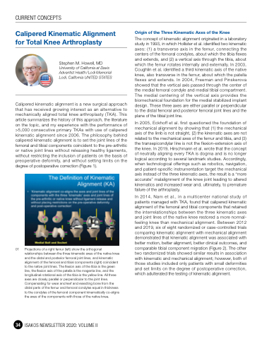
01 Projections of a right femur (left) show the orthogonal relationships between the three kinematic axes of the native knee and the distal and posterior femoral joint lines, and kinematic alignment of the femoral and tibial components (right) coincident to the native joint lines. The flexion axis of the tibia is the green line, the flexion axis of the patella is the magenta line, and the longitudinal rotational axis of the tibia is the yellow line. All three axes are closely parallel or perpendicular to the joint lines. Compensating for wear and kerf and resecting bone from the distal parts of the femur and femoral condyles equal in thickness to the condyles of the femoral component kinematically co-aligns the axes of the components with those of the native knee.
34 ISAKOS NEWSLETTER 2020: VOLUME II
