
Page 21 - ISAKOS 2021 Newsletter Volume 1
P. 21
The Osteochondral Lesion: Management Options Now and Going Forward
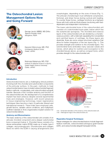
morphologies, depending on the zone of tissue (Fig. 1). The articular chondral layer must withstand compressive, frictional, and sheer forces during cyclical joint loading. To counter these forces, cartilage functions as a biphasic tissue with both solid and fluid phases functioning with fluid- dependent and independent mechanisms.
The lower subchondral bone of the osteochondral unit consists of a subchondral bone plate, below which lies the subarticular spongiosa. The chondral and osseous layers of the osteochondral unit are divided by a chondro- osseous junction, with a tidemark separating the noncalcified and calcified layers of cartilage. As these layers are dissimilar, there are some unique structural variations that allow for better integration of the two layers (Fig. 1). This provides strength and nutrition to the chondral layers. The subchondral bone embodies many vascular canals and nerves, which allow for nutrition and nociception to the chondral tissues above, as well as compact bone, which provides strength to the osteochondral unit.
01A Schematic Illustration of the anatomy of the osteochondral unit.
01B Histological image of the osteochondral unit.
Reparative Surgical Techniques
Repair strategies for osteochondral lesions include fragment- fixation methods, osteochondral autologous graft transfer (OAT), and osteochondral allograft transplant (OCA).
Introduction
George Jacob, MBBS, MS Ortho
Tejasvini Hospital, Kadri, Mangalore, INDIA
Kazunori Shimomura, MD, PhD
Hoshigaoka Medical Center, Osaka, JAPAN
Norimasa Nakamura, MD, PhD
Institute for Medical Science in Sports, Osaka Health Science University, Osaka, JAPAN
Osteochondral lesions are a challenging clinical problem involving both the chondral and subchondral components of the articular surface. In the past, methods to treat osteochondral lesions have included osteochondral fragment fixation methods, mosaicplasty, and osteochondral autograft transfer. Because of the complex anatomy and function of the osteochondral unit, the development of a comprehensive implant has been arduous despite several experimental trials and strategies. However, with recent advances in tissue engineering technology, newer regenerative techniques have become a topic of particular interest and translational research. These trials have employed cells, growth factors, variable culture conditions, and multiphasic scaffold materials to find a regenerative implant solution for osteochondral defects.
Anatomy and Biomechanics
The basic anatomy of the osteochondral unit consists of an articular chondral component and a deeper subchondral bone component below it. Chondral tissue consists of a dense extracellular matrix (ECM) made up of water, type-II collagen, and proteoglycans. Chondrocytes and collagen fibers lie within the ECM in varying arrangements and
CURRENT CONCEPTS
ISAKOS NEWSLETTER 2021: VOLUME I 19
