
Page 22 - ISAKOS 2021 Newsletter Volume 1
P. 22
CURRENT CONCEPTS
The Osteochondral Lesion: Management Options Now and Going Forward
Each of these techniques has its own strengths and weaknesses, and outcomes are dependent on patient and lesion-specific variables. Table I summarizes the reparative techniques available for osteochondral lesions.
Table I. Summary of Reparative Techniques for Osteochondral Lesions
Technique Weaknesses
Regenerative Techniques
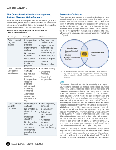
Regenerative approaches for osteochondral lesions have been challenging, and strategies have utilized cells, growth factors, and various scaffold materials. The solution must result in a hyaline cartilage layer supported by a material to emulate subchondral bone, and, most importantly, both materials should integrate with each other. This is the reason for the development of multiphasic scaffolds. The ideal attributes of a regenerate osteochondral unit are highlighted in Figure 2.
02 The ideal attributes of an osteochondral implant. The two layers of the implants including the chondral and subchondral areas require different properties but both must have bioactivity and mechanical strength.
Cells
Cells accomplish and mediate the bioactivity of an implant. Sources have included autologous cell biopsies as well as stem cells, and each source has its own advantages and challenges. Autologous chondrocyte biopsy was popular but lacked sufficient cell numbers. This led to in vitro expansion being advocated, which resulted in cell dedifferentiation and loss of chondrogenic phenotype. Among stem cells, the available cell types are embryonic stem cells (ESCs) and mesenchymal stem cells (MSCs); however, given the ethical obstacles associated with ESCs, MSCs have been preferred. MSCs have been isolated from several tissue sources, with bone marrow, adipose tissue, and synovium being the most popular. Among MSCs, synovium-derived MSCs are known to be superior in terms of chondrogenic and osteogenic differentiation but require considerable in vitro cell expansion for clinical use. MSCs exhibit paracrine functions, which promote cell proliferation, stimulate anti-inflammatory processes, and encourage endogenous cell recruitment, improving regenerate quality and implant integration.
The recent discovery of induced pluripotent stem (iPS) cells may allow for a new cell source. iPS cells such as ESCs have limitless proliferative potential and superior differentiation capacity, without the ethical drawbacks of ESCs, but are similar in that teratoma formation is a risk factor.
Strengths
Osteochondral fragment fixation
Osteochondral autologous graft transfer
• Limited quantity
• Donor-site morbidity
• Possible >1 surgical site
• Intraoperative decision
possible
• Mature hyaline cartilage
• No immune reaction
• Perfect size and contour if reduced properly
• Fragment may not be viable
• Dependent on fragment size, integrity, and time from injury
• Implant required
• Possible implant removal
• Tissue reaction
Osteochondral allograft transplant
• Mature hyaline cartilage
• No Immune reaction
• Intraoperative decision
possible, with no graft availability concerns
• Addresses subchondral and chondral layer
• Mature hyaline cartilage
• No limitation in size of donor graft
• Immediate defect fill
• No donor-site morbidity
• Addresses subchondral and chondral layer
• Immunogenicity concerns
• ↓ cell viability
• Difficult to procure and store
• Additional expense
• Possible graft size mismatching
20 ISAKOS NEWSLETTER 2021: VOLUME I
