
Page 19 - ISAKOS 2021 Newsletter Volume 1
P. 19
The arthrotomy commences with the creation of a square synovial flap (Fig. 7). The posterior limit is vertical, just anterior to the MCL and distal to the medial meniscus. The geniculate artery is coagulated in its wall.
Then, with gentle retration on the PT at its medial distal insertion, the medial patellar tibial ligament (MPTL) is detached till the PT fibers are well individualized.
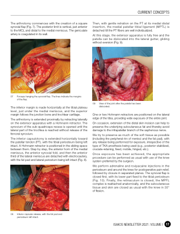
At this stage, the extensor apparatus is fully free and the patella can be dislocated into the lateral gutter, gliding without eversion (Fig. 9).
09 View of the joint after the patella has been dislocated.
One or two Hohmann retractors are positioned on the lateral edge of the tibia, providing wide exposure of the entire joint.
On occasion, extension of the distal skin incision can help to preserve the underlying subcutaneous fat and thereby avoid damage to the infrapatellar branch of the saphenous nerve.
We try to preserve as much of the soft tissue as possible (including the peripheral rim of menisci and the fat pad), with any release being performed for exposure, irrespective of the type of TKA prosthesis being used (e.g., posterior stabilized, cruciate-retaning, fixed, mobile, hinged, etc.).
Once exposure has been achieved, the appropriate procedure can be performed as usual with use of the knee system preferred by the surgeon.
We perform adrenaline and rovipacaine injections in the periosteum and around the knee for postoperative pain relief, followed by closure in separated planes. The synovial flap is closed first, with its lower part fixed to the tibial periosteum (Fig. 10). Finally, the retinaculum is closed, the MPFL complex is reattached anatomically, and the subcutaneous tissue and skin are closed as usual with the knee in 30° of flexion.
07 Forceps hanging the synovial flap. The lines indicate the margins of the flap.
The inferior margin is made horizontally at the tibial plateau level, just under the medial meniscus, and the superior margin follows the junction bone and trochlear cartilage.
The arthrotomy is extended proximally by retracting laterally on the extensor apparatus with a Hohmann retractor. The synovium of the sub-quadriceps recess is opened until the lateral part of the trochlea is reached without release of the femoral synovium.
The inferior capsulotomy is extended horizontally toward the patellar tendon (PT), with the tibial periosteum being left intact. A Hohmann retractor is positioned in the sliding space between them. Step by step, the anterior horn of the medial meniscus, the anterior synovial fold, and then the anterior third of the lateral meniscus are detached with electrocautery, with the fat pad and lateral periostum being left intact (Fig. 8).
CURRENT CONCEPTS
08 Inferior capsular release, with the fat pad and periosteum left intact.
ISAKOS NEWSLETTER 2021: VOLUME I 17
