
Page 31 - ISAKOS 2018 Winter Newsletter
P. 31
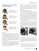
 Perspectives on Instrumented Pivot-Shift Testing
Several instrumented measurement systems have been developed in an attempt to achieve comparable and meticulous measurements with use of the pivot-shift test. Some of these systems can be used in a non-invasive manner and thus are clinically applicable. Three major examples of instrumented pivot-shift test systems are described below.
Electromagnetic Measurement System
In vivo knee kinematics during the pivot-shift test were first evaluated with use of an electromagnetic system by Bull et al. in 2002, with the electromagnetic sensors being fixed to the bones with pins. The disadvantage of that method was that it could only be used intraoperatively. We then developed a noninvasive method for the application of a similar electromagnetic system (Liberty, Polhemus, Colchester, VT, USA) in order to allow for wider clinical use. With that method, electromagnetic sensors are attached over the femur and tibia with use of plastic straps and a third sensor is used to input the 3D positions of anatomic osseous landmarks for both bones (Fig. 1).
An anatomical coordinate system for the knee is configured in a virtual space on the basis of the 3D positional relationship of the two bones, and knee kinematics are recorded in 6 degrees of freedom during the pivot-shift test (Fig. 2). Anterior tibial translation and tibial acceleration are calculated from the data, and these parameters are used when determining the clinical grading of the results of the pivot-shift test.
01 02
This electromagnetic system is now commercially available with a user-friendly console that includes original software to automatically calculate measurement parameters (Joint Instability Measurement Interface JIMI Kobe, Arthrex Japan, Tokyo, JAPAN) (Fig. 3), although its sales area is currently limited to Japan.
01 The 3D position of the anatomic landmark is digitized with use of a stylus equipped with an electromagnetic receiver. Seven anatomic landmarks are registered to configure the knee coordinate system.
02 The pivot-shift test is performed while evaluating 6-degrees-of-freedom knee kinematics with use of the electromagnetic system. Tibial anterior translation and acceleration are calculated for further analysis.
Introduction
Yuichi Hoshino, MD, PhD Department of Orthopaedic Surgery, Graduate School of Medicine,
Kobe University, Kobe, JAPAN
Daisuke Araki, MD, PhD Department of Orthopaedic Surgery, Graduate School of Medicine,
Kobe University, Kobe, JAPAN
Takehiko Matsushita, MD, PhD Department of Orthopaedic Surgery, Graduate School of Medicine,
Kobe University, Kobe, JAPAN
Ryosuke Kuroda, MD, PhD Department of Orthopaedic Surgery, Graduate School of Medicine,
Kobe University, Kobe, JAPAN
There is still considerable variability in the knee-laxity evaluations performed in research studies on the anterior cruciate ligament (ACL). Quantitative evaluation of anterior laxity is conducted almost exclusively with use of KT-1000 measurements, whereas rotational laxity is assessed only with use of the manually performed pivot-shift test1. It is well known that the manual pivot-shift test is very subjective and provides only 4 levels of grading. Therefore, subtle differences in rotational laxity cannot be consistently detected with the manual pivot-shift test.
The current problem is that residual rotational laxity following ACL reconstruction is not always detectable with the manual test and does not consistently produce sizable improvement of rotational laxity. Quantitative evaluation of the pivot-shift test is therefore necessary in order to improve the ability to assess the results of ACL reconstruction.
CURRENT CONCEPTS
ISAKOS NEWSLETTER 2018: VOLUME I 29
