
Page 32 - ISAKOS 2018 Winter Newsletter
P. 32
CURRENT CONCEPTS
Perspectives on Instrumented Pivot-Shift Testing
Triaxial Accelerometer System
As noted in our experimental study on the electromagnetic system, an acceleration surge is observed during the pivot- shift test. Direct measurement of this tibial acceleration has been shown to be feasible with use of the KiRA system (Kinematic Rapid Assessment; Orthokey LLC, Lewes, DE, USA).
04
With that system, an accelerometer is attached over the lateral aspect of the shank between the tibial tuberosity and Gerdy’s tubercle with use of a wrapping band and is wirelessly connected to a computer equipped with Bluetooth, and the acceleration of knee movement is recorded with the original software. KiRA is also commercially available worldwide now, but an official broker is commonly required to import such a high-technology device in most countries outside of the EU.
Clinical Usability of Noninvasive Quantitative Measurement Devices
These 3 instrumented systems for the pivot-shift test seem to be feasible in our clinical practice, but their diagnostic accuracy has not been compared. Therefore, we conducted a study to compare these 3 systems in terms of the clinical detection of ACL deficiency. The pivot-shift test was performed for 30 patients with unilateral ACL injuries who were under general anesthesia. Three instruments (the accelerometer system [KiRA], the image-analysis system involving the using iPad [iPad], and the electromagnetic measurement system [EMS]) were used simultaneously to evaluate 2 parameters: tibial acceleration (as monitored with the KiRA and EMS systems) and tibial anteroposterior translation (as recorded with the iPad and EMS systems).
Although all 3 systems could detect ACL deficiency, receiver operator characteristic (ROC) curve analysis demonstrated that the EMS method was more accurate than the KiRA and iPad methods for the detection of ACL insufficiency.
Simple Image Analysis with Use of an iPad
The pivot-shift phenomenon was first reported as the anterior subluxation of the lateral tibial plateau and its spontaneous reduction. However, as some ACL-deficient patients show visible abnormal tibial translation on the lateral side of the knee joint during the test, it was hypothesized that lateral compartment translation may have an effect on the grading of the test results. Therefore, a trial was performed to capture the surface movement during the pivot-shift with use of a digital camera and subsequent image analysis with a computer. Three round stickers, 0.75 inch in diameter (Color Coding Labels; Avery Dennison, Pasadena, CA, USA), were attached to the skin over specific osseous landmarks (the lateral epicondyle, Gerdy’s tubercle, and the fibular head) to allow for image analysis. A video capturing the lateral aspect of the knee joint was made during the pivot-shift test with the digital camera. The video was then processed in the computer to evaluate the anteroposterior movement of the tibial points (Gerdy’s tubercle and the fibular head) relative to the femoral point (the lateral epicondyle). The anteroposterior tibial translation on the lateral side of the knee joint was successfully captured and was found to be related to the clinical grading of the results of the test.
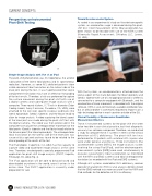
The iPad (Apple, Cupertino, CA, USA) has the capacity to capture video and to perform image analysis at the same time. Therefore, we designed a specially programmed iPad application for pivot-shift image analysis (PIVOT, Impellia, Pittsburgh, PA, USA) (Fig. 4).
The iPad application will be able to be purchased and downloaded anywhere in the world, once it arrives in the online store. The PIVOT application is currently under preparation for sale.
03
30 ISAKOS NEWSLETTER 2018: VOLUME I
