
Page 29 - ISAKOS Newsletter 2016 Volume 1
P. 29
CURRENT CONCEPTS
Clinical Relevance of Intraoperative Soft Tissue Balance Assessment
Considering clinical significance of intraoperative assessment, we should confirm that intraoperative values assessed with the tensor, reflects the postoperative soft tissue balance. Hence, we investigated the correlation between the intraoperative values assessed with the tensor and the 5-year postoperative values assessed with stress radiographs at extension and flexion. In CR TKA, postoperatively both the joint component gap and ligament balance at extension and flexion showed positive correlations with the intraoperative values of 10 degrees of flexion and 90 degrees of flexion. However, in PS TKA, whereas postoperatively both the joint component gap and ligament balance at extension showed positive correlation with intraoperative values of 10 degrees of flexion, postoperatively neither joint component gap nor ligament balance at flexion showed correlation with that at 90 degrees of flexion.
These results indicate that the intraoperative measurements of soft tissue balance by the tensor reflect postoperative values assessed by the stress radiographs even at the 5-year follow-up.
However, despite existing correlations in extension, there were no correlations in flexion in both the joint component gap and ligament balance between intra and postoperative values in PS TKA. This discrepancy in PS TKA may be caused by flexion instability due to a larger flexion gap compared to extension gap.
Acquisition of high flexion angle after TKA is one of the factors leading to patient satisfaction. Therefore, we focused on the postoperative flexion angle in relation to intraoperative soft tissue balance. In the series of studies in PS TKA, joint gap change value (90-0o) with PF joint reduced, not everted, showed inverse correlation with the postoperative knee flexion angle and posterior condylar offset. However, in another series of studies on CR TKA, the postoperative flexion angle was positively correlated with the joint gap change value (90-0°). In either case, multivariate regression analysis among various values including joint gap change values, ligament balance, and pre-operative knee flexion angle, demonstrated that the preoperative knee flexion angle and the joint gap change value (90-0°) had a significant independent effect on the postoperative knee flexion angle. One of the reasons for this discrepancy may be the different patterns of soft tissue balance between PS and CR TKA. In that report, CR TKA in comparison to PS TKA, showed significantly smaller gaps when the arc of movement ranged from mid-to deep flexion. The posterior cruciate ligament (PCL) in the osteoarthritic knee is considered relatively rigid and shortened, despite being relatively macroscopically intact. When we consider the flexion gap tightness, Ritter et al reported that 30% of CR TKA required ligament balancing to obtain a smooth flexion arc.
If the PCL was too tight, excessive femoral rollback resulted in anterior lift-off of the tibial trial in flexion, leading to limitation of flexion. To improve postoperative flexion, balancing the flexion gap can result in a satisfactory range of motion. In our series of studies on CR TKA, it was identified that 16% more flexion gap tightness (smaller flexion gap than extension gap) resulted in a smaller flexion angle. Similarly, using a commercially available knee balancer with the measurement under 80 N distraction force, Higuchi et al reported that flexion medial / lateral gap tightness led to restriction of the flexion angle. Therefore, in these cases, surgeons are advised to avoid flexion gap tightness by soft tissue release such as PCL.
02
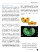
02 Verasense Knee System (OrthoSensor Inc., Dania Beach, Florida) Verasense Knee System provides two essential profiles; dynamic Femoral Contact Points enabling the surgeon to visualize femoral component rollback and tibial tray rotation to improve tibial-femoral articulation; and Kinetic TrackingTM feature displaying femoral component motion paths in the medial and lateral compartments to enable a full kinetic (force and motion) assessment of knee function.
ISAKOS NEWSLETTER 2016: Volume I 27
