
Page 28 -
P. 28
CURRENT CONCEPTS
Femoral Tunnel Placement in ACL Reconstruction Central Footprint vs. AM Bundle
Mark Clatworthy, FRACS
Middlemore Hospital Auckland, NEW ZEALAND
Additional Authors:
Andrew Pearle, MD
HSS, New York, USA
Andrew Williams, MB BS, FRCS, FRCS (Orth), FFSEM(UK) Fortius Clinic, London, UK
Martin Lind, Prof., MD, PhD
Aarhus University Hospital, DENMARK
Introduction
‘Anatomic’ ACL reconstruction, implying a central femoral footprint tunnel position, has been advocated to improve knee stability, function and outcome. Many surgeons changed their surgical technique from a transtibial approach which typically results in a high AM femoral tunnel and posterior tibial tunnel placement to a transportal technique whereby the femoral tunnel position can be determined independent of tibial tunnel position. Many continued placing the femoral tunnel in the AM bundle position, but more recently, a central so-called ‘anatomic’ position was advocated.
This choice of femoral tunnel was based on anatomical and time zero biomechanical studies which suggested better control of rotational instability. It was predicted that more ‘anatomical’ placement would result in a fewer failures and reduced incidence of post ACL reconstruction osteoarthritis.
The implication of the term ‘anatomic’ is that the more traditional AM bundle tunnel position in the femur is non-anatomic. However, the AM position is still within the anatomic footprint; it is eccentrically located in the AM portion of the footprint rather then centralized within the footprint. Therefore, an AM position should still be considered an anatomic position.
A number of high volume surgeons noticed a higher ACL graft rupture rate with a central femoral tunnel position. This has resulted in a revisitation of ACL anatomy, biomechanical studies and clinical studies to determine the reasons for these findings.
This review article will detail the newer anatomical and histologic studies which show a direct and indirect ACL origin, biomechanical analysis of the direct and indirect fibres which show most of the load goes through the direct fibres, and recently completed clinical studies evaluating the effect of moving to a central femoral tunnel position on ACL graft failure
Anatomy & Biomechanics
ACL fibers form an oblong footprint that are organized such that the midsubstance of the ACL is narrower than the femoral footprint. Anatomic dissections have demonstrated that while the femoral footprint is oval, the native ACL forms a flat, ribbon like structure with a width of 9 – 16 mm and a thickness of only 2 – 4 mm as it takes off from the bone.
26 ISAKOS NEWSLETTER 2015: Volume II
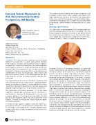
Direct
Indirect
Histologic analysis of the femoral footprint has provided further clarification of the relationship of the footprint with functional aspects of the native ACL. The femoral origin of the ACL has distinct direct and indirect insertions, which were shown by histology and 3-dimensional volume- rendered computed tomography. The direct insertion consisted of dense collagen fibers anterior in the femoral footprint connected to a bony depression immediately posterior to the lateral intercondylar ridge. These direct fibers extended an average of 5.3 ± 1.1 mm posteriorly but do not continue to the posterior femoral articular cartilage. The indirect insertion consisted of more posterior collagen fibers that extend to blend into the articular cartilage of the posterior aspect of the lateral femoral condyle. This membrane-like tissue was located between the direct insertion and the posterior femoral articular cartilage, and had an average width of 4.4 ± 0.5 mm in the anteroposterior direction on average. This anterior band of ACL tissue with the direct insertion histologically corresponded to the fibers in the anterior, more isometric region of the femoral footprint. Conversely, the more posterior band of fibers, that have an indirect insertion histologically, corresponded to the more anisometric region and are seen macroscopically as a fan like projection extending to the posterior articular cartilage.
01
