
Page 27 - ISAKOS 2018 Winter Newsletter
P. 27
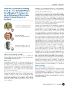
 Talar Osteoperiosteal Grafting From the Iliac Crest (TOPIC): A Novel Surgical Technique for Large Primary and Secondary Osteochondral Defects of
In patients with large defects and those who have had a failure of first-line surgical treatment, more extensive and invasive surgery may be necessary.
For those specific clinical indications, treatment strategies that are frequently employed include osteochondral allograft procedures, osteochondral autograft transfer system (OATS) procedures (involving either a single plug or mosaicplasty), and classic and matrix-associated chondrocyte implantation (ACI, MACI). Each of these options has clinical advantages and disadvantages. Allografts, for instance, have a high failure rate: in a number of studies, graft rejection was observed as a result of immunological reactions.
Although the OATS procedure is also regarded as an effective surgical option, with a 90% success rate reported in the literature, donor-site morbidity rates have been reported to range from 11% to 35% when the graft was harvested from the ipsilateral femoral condyle. Furthermore, the graft that is harvested from the femoral condyle frequently does not properly restore the surface of the talar dome, resulting in incomplete incorporation of the implanted graft as well as bone cyst formation. Previously, a new technique involving the use of autologous osteoperiosteal cylinder grafts was described by Hu et al. and Chen et al. The surgical treatment yielded promising results; however, the downside of that method is that the size of the cylinder autograft is static and is determined preoperatively, thereby compromising optimal individualized treatment for large talar osteochondral defects.
In order to address these disadvantages, we developed a new surgical technique for the treatment of large talar osteochondral defects or secondary lesions: the Talar Osteoperiosteal Grafting from the Iliac Crest (TOPIC) procedure. With this press-fit technique, the autograft is harvested from the ipsilateral iliac crest, a site that has been shown to have chondrogenic potential because the cambium layer of periosteum contains chondrocyte precursor cells4. A bone-periosteal transplant for bone-cartilage repair meets the 3 requirements for tissue engineering: a source of cells, a scaffold, and local growth factors. In addition, the curvature of the iliac crest is highly similar to the curvature of the talar bone. In this Pearls & Pitfalls section, we will describe the surgical technique for the newly developed TOPIC procedure.
Surgical Technique
Step 1: Medial Malleolar Osteotomy
The TOPIC procedure is carried out with the patient under general or spinal anesthesia following the administration of antibiotic prophylaxis. The patient is placed in the supine position with a tourniquet applied around the proximal aspect of the leg. The team prepares for surgery and carries out a time-out procedure, after which the entire ipsilateral lower leg is prepared and draped.
the Talus
Gino M.M.J. Kerkhoffs, MD, PhD
Amsterdam, THE NETHERLANDS
Jari Dahmen, BSc
Amsterdam, THE NETHERLANDS
Sjoerd A.S. Stufkens, MD, PhD
Amsterdam, THE NETHERLANDS
Pearls & Pitfalls–Surgical Technique
Talar Osteoperiosteal Grafting From the Iliac Crest (TOPIC): A Novel Surgical Technique for Large Primary and Secondary Osteochondral Defects of the Talus
The Concept
Osteochondral defects of the talus are characterized by damage to the talar articular cartilage and subchondral bone. Such defects may be due to an ankle fracture or sprain, an avascular episode, and, possibly, a genetic predisposition. These injuries have a severe impact on the quality of life of active patients as a result of deep ankle pain during weight- bearing and sporting activities.
The initial treatment of these defects typically consists of conservative measures, after which—in cases of persistent symptoms—surgical therapy can be considered. First-line surgical treatment options that are frequently applied include arthroscopic debridement of the defect, bone marrow stimulation, fixation, and retrograde drilling when cystic lesions are present.
CURRENT CONCEPTS
ISAKOS NEWSLETTER 2018: VOLUME I 25
