
Page 35 - ISAKOS 2019 Newsletter Vol II
P. 35
Distal Biceps Endoscopy: All-Endoscopic Exploration, Repair, and Reconstruction of Distal Biceps Tears
Deepak N. Bhatia, MS(Orth), DNB(Orth)
Sportsmed Mumbai
Mumbai, India
Introduction
Distal biceps endoscopy is a minimally invasive diagnostic and therapeutic technique for the management of distal biceps tendon (DBT) pathology. The endoscopic technique has evolved from a single-incision diagnostic technique to an endoscopic-assisted and all-endoscopic technique for DBT repair and reconstruction1,2. The advantages and potential pitfalls have been identified in cadaveric and clinical studies1, 2 – 5, and knowledge of the principles and technical variations of biceps endoscopy is necessary prior to the transition from open to endoscopic procedures around the distal biceps.
Surgical Anatomy, Portal Safety, and Technical Feasibility
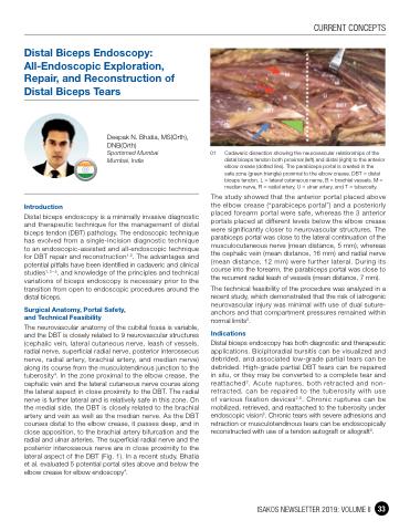
The neurovascular anatomy of the cubital fossa is variable, and the DBT is closely related to 9 neurovascular structures (cephalic vein, lateral cutaneous nerve, leash of vessels, radial nerve, superficial radial nerve, posterior interosseous nerve, radial artery, brachial artery, and median nerve) along its course from the musculotendinous junction to the tuberosity4. In the zone proximal to the elbow crease, the cephalic vein and the lateral cutaneous nerve course along the lateral aspect in close proximity to the DBT. The radial nerve is further lateral and is relatively safe in this zone. On the medial side, the DBT is closely related to the brachial artery and vein as well as the median nerve. As the DBT courses distal to the elbow crease, it passes deep, and in close apposition, to the brachial artery bifurcation and the radial and ulnar arteries. The superficial radial nerve and the posterior interosseous nerve are in close proximity to the lateral aspect of the DBT (Fig. 1). In a recent study, Bhatia et al. evaluated 5 potential portal sites above and below the elbow crease for elbow endoscopy4.
01 Cadaveric dissection showing the neurovascular relationships of the distal biceps tendon both proximal (left) and distal (right) to the anterior elbow crease (dotted line). The parabiceps portal is created in the
safe zone (green triangle) proximal to the elbow crease. DBT = distal biceps tendon, L = lateral cutaneous nerve, B = brachial vessels, M = median nerve, R = radial artery, U = ulnar artery, and T = tuberosity.
The study showed that the anterior portal placed above the elbow crease (“parabiceps portal”) and a posteriorly placed forearm portal were safe, whereas the 3 anterior portals placed at different levels below the elbow crease were significantly closer to neurovascular structures. The parabiceps portal was close to the lateral continuation of the musculocutaneous nerve (mean distance, 5 mm), whereas the cephalic vein (mean distance, 16 mm) and radial nerve (mean distance, 12 mm) were further lateral. During its course into the forearm, the parabiceps portal was close to the recurrent radial leash of vessels (mean distance, 7 mm).
The technical feasibility of the procedure was analyzed in a recent study, which demonstrated that the risk of iatrogenic neurovascular injury was minimal with use of dual suture- anchors and that compartment pressures remained within normal limits5.
Indications
Distal biceps endoscopy has both diagnostic and therapeutic applications. Bicipitoradial bursitis can be visualized and debrided, and associated low-grade partial tears can be debrided. High-grade partial DBT tears can be repaired in situ, or they may be converted to a complete tear and reattached2. Acute ruptures, both retracted and non- retracted, can be repaired to the tuberosity with use of various fixation devices2,5. Chronic ruptures can be mobilized, retrieved, and reattached to the tuberosity under endoscopic vision3. Chronic tears with severe adhesions and retraction or musculotendinous tears can be endoscopically reconstructed with use of a tendon autograft or allograft3.
CURRENT CONCEPTS
ISAKOS NEWSLETTER 2019: VOLUME II 33
