
Page 34 - ISAKOS 2019 Newsletter Vol II
P. 34
CURRENT CONCEPTS
In Vitro-Generated Autologous Bone Graft: Progress Toward a Desired Surgical Tool to Assist in Reconstruction and Repair
A Glimpse Toward the Future
Nahum Rosenberg, MD, FRCS, Prof.
Technion -IIT
Haifa, Israel
The problem: nonunion and slow healing of bone defects. The solution: a bioengineered bone graft product.
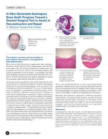
01
01A Photomicrograph showing
the bone spicules generated
in vitro, originating from osteoblasts cultured as explant culture from bone marrow (hematoxylin and eosin).
01C Photomicrograph showing a critical bone gap in rat calvarium, 6 weeks after
the creation of the gap (hematoxylin and eosin). No evidence of bone bridging of the gap exists.
01B Bone-like material generated in vitro.
Bone has a high potential to regenerate after damage. However, large defects with inadequately vascularized bone gaps may not achieve satisfactory healing and may progress to nonunion. About 10% of these patients will require implantation of bone grafts1. Cumulatively, 500,000 bone-grafting procedures are performed annually in the U.S. alone2, and the demand for bone grafts is expected to rise as the population ages and life expectancy increases1.
Autologous bone-grafting is the gold standard for the reconstruction of critical bone defects. However, autologous grafts have several disadvantages, such as donor-site morbidity and limited availability. The use of alternatives such as allografts or xenografts is limited because of the risks of rejection, infection, and nonunion. Progress in bone tissue engineering is essential in order to provide an unlimited source of autologous bone for grafting. Fortunately, the method of generating autologous bone-like material, originating from patient-specific osteoblast-like cells grown from bone marrow on a -tricalcium phosphate-supporting 3-dimensional matrix under optimal biomechanical conditions, already exists3. The generated live-bone material might be advantageous over existing inorganic fillers, which have low osteoconductivity and osteoinductivity, as well as over cell-therapy products, which have an uncertain ability to proceed to bone generation after implantation. This newly developed tissue-engineering method showed promising results in initial animal studies (Fig. 1).
01D Hotomicrograph showing bridging of the critical bone gap by young woven bone in rat calvarium, 6 weeks after the implantation of in vitro- generated bone.
32 ISAKOS NEWSLETTER 2019: VOLUME II
Thus, the methodology for in vitro live human bone generation already exists, and we should anticipate that this source of autologous bone for grafting, which does not have the disadvantages of surgical morbidity and limitation of the volume of the graft, is ready to be developed for clinical use.
Naturally, the method is expected to fulfill the regulatory requirements, including clinical studies, and this process might take several years. However, on the basis of the promising initial preclinical evidence, the musculoskeletal surgeon community should expect an important breakthrough in bone graft use in the future.
References
1. Alexander PG, Hofer HR, Clark KL, Tuan RS: Mesenchymal Stem Cells in Musculoskeletal Tissue Engineering. In Principles of Tissue Engineering. 4th edition. Edited by Lanza R, Langer R, Vacanti J. Waltham, MA, USA: Elsevier; 2014: 1171-1200 2. Mauffrey C, Madsen M, Bowles RJ, Seligson D: Bone graft harvest site options in orthopaedic trauma: a prospective in vivo quantification study. Injury 2012, 43:323-326. 3. Rosenberg N, Rosenberg O. Extracorporeal human bone-like tissue generation. Bone and Joint Research (BJR) 2012; 1:1-7
