
Page 25 -
P. 25
05
The Rotator Cuff (Fig. 5) The infraspinatus has been detached to expose the capsule. (Courtesy Hiro Sugaya)
06
Intra-muscular rotator cuff tendons (Fig. 6). The subscapularis has 4 tendons, the supraspinatus is a bipennate muscle, with 1 tendon; and the infraspinatus has an oblique head (head depressor) and transverse head (external rotation). (Created by Dr. Afsana Hasan).
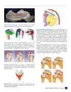
07 Rotator Cuff Tears (Fig. 7); Crescent, L-shaped, reverse L-shaped tears have a different appearance and require different methods of mobilization and fixation. (Courtesy of
Steve Burkhart).
08
Deltoid (Fig. 8), There are 7 segments derived from the anterior, lateral and posterior, acromion. (Courtesy of Eiji Itoi, modified from Rispoli et al).
09 Functional glenoid labrum (Fig. 9). The inferior labrum is a fixed convex bumper mounted on the osseous glenoid. The superior labrum is a mobile organ of tension, which attaches “off the glenoid face.” Three is a synovial lined cleft between the labrum and the glenoid, which allows it to adapt its shape in various positions. The superior labrum is actively controlled by the biceps tendon, and passively controlled by the SGHL and MGHL at the extremes of motion. The dynamic biceps, rotator cuff and deltoid all compress the humeral head onto the “static” inferior glenoid and labrum, to provide joint
stability.
AC Joint Instability (Fig. 10); Pattern and classification of progressive and sequential AC joint instability and scapula medicalization. Stage 1 – Failure of AC ligaments and capsule. Stage 2 – Failure of the trapezoid at the coracoid. Stage 3 – Failure of the conoid at the clavicle. Stage 4 – Stripping of the inferior periosteum from the medial clavicle. (Concept Greg Bain, Image Copyright Dr. Martin Langer).
10
ISAKOS NEWSLETTER 2015: Volume II 23
