
Page 37 - ISAKOS 2019 Newsletter Vol II
P. 37
CURRENT CONCEPTS
03 Endoscopic images showing a normal DBT (upper image), a partially torn tendon (lower left image) and completely torn non-retracted tendon (lower right image). T = tuberosity, Pr = probe.
Complete tears demonstrate a bare tuberosity, and remnants of the tendon stump may be visualized (Fig. 3). A grasper is used to deliver the proximal end of the non-retracted DBT via the distal anterior portal under endoscopic visualization. DBT tears that have retracted proximal to the elbow crease are explored and retrieved via the mid-biceps portal, which is placed approximately 1 to 2 cm proximal to the proximal parabiceps portal in the midline of the forearm. Chronic retracted tears are released to achieve adequate tendon length; inadequate length, degenerated tendons, or musculotendinous tears are reconstructed with use of hamstring autograft tendon. The tendon is then shuttled across the elbow crease and is retrieved via the distal anterior portal. The bicipital tuberosity is prepared with use of a 3-mm shaver and burr, and two double-loaded all-suture anchors (Suturefix Ultra, 1.9 mm; Smith & Nephew, USA) are passed 1 cm apart (Fig. 4). One suture from each pair is used to whipstitch the tendon, and the other end is pulled to dock the tendon on the tuberosity. Non-sliding knots are used to fix the DBT securely to the tuberosity. Alternately, button devices (Endobutton [Smith & Nephew, USA] or BicepsButton [Arthrex, USA]) may be used for DBT fixation (Fig. 5). Dynamic assessment is used to evaluate tendon-bone contact and fixation.
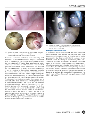
04 Endoscopic images showing the placement of double-loaded all-suture anchors (AN) into the tuberosity, approximately 1 cm apart (arrows). T = tuberosity.
Postoperative Rehabilitation
A bulky soft-tissue dressing with the elbow in 90° of flexion is used in the immediate postoperative period, and this dressing is changed to portal dressing on the fourth postoperative day. Sling immobilization is necessary for up to 4 to 6 weeks, depending on the chronicity of the tear. Thereafter, a hinged elbow brace is used for controlled range-of-motion, and active-assisted and active range- of-motion exercises are initiated and continued for 2 to 4 weeks. Next, low-intensity muscle strengthening exercises are started, and the patient is permitted to resume all activities of daily living. Biceps and triceps strengthening is begun at 12 weeks and is progressively increased over the next 3 months. Heavy-lifting work and sports are permitted after 4 to 6 months.
ISAKOS NEWSLETTER 2019: VOLUME II 35
