
Page 27 - ISAKOS 2019 Newsletter Vol II
P. 27
Technical Tips for Simultaneous ACL Surgery with HTO
Physical examination reveals pain on palpation that is localized to the medial compartment, and a variety of tests are used to detect instability. ACL injury or failure of reconstruction can be seen on magnetic resonance imaging (MRI), degeneration of the joint can be seen on standing radiographs, and varus malalignment can be seen on standing long-leg radiographs. The amount of correction is a matter of surgeon preference; some surgeons aim for a neutral mechanical axis, whereas others aim for slight overcorrection of the mechanical axis to the Fujisawa point2. In general, a neutral mechanical axis may be preferable for younger patients who would like to return to sports.
Patient Positioning
After the induction of general or regional anesthesia and intravenous administration of prophylactic antibiotics, the patient is placed in supine position on a radiolucent table, with both knees hanging down from the table with the knees flexed at 90°. A tourniquet is placed at the proximal part of the thigh, and a lateral post is placed at thigh level. The C-arm imaging device is placed on the lateral side of the knee because the surgeon will be operating on the medial side during the osteotomy.
Surgical Steps
1 Graft harvest
2 Arthroscopy (examination, chondral and meniscal
interventions, debridement)
3 Femoral tunnel drilling
4 Medial opening-wedge osteotomy (with preliminary fixation) 5 Tibial tunnel drilling
6 Graft passage
7 Definitive fixation of osteotomy
8 Tensioning and fixation of the graft
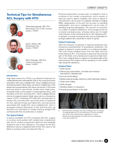
01 Anteroposterior radiograph (A), lateral radiograph (B), and sagittal T2-weighted MRI scan (C) of the left knee of 35-year-old patient with an ACL injury that had been neglected for 10 years. Standing long-leg radiograph showing both lower extremities
of the same patient (D).
Introduction
Tahsin Beyzadeoglu, MD, Prof.
Beyzadeoglu Clinic & Halic University Istanbul, TURKEY
Kerem Yildirim, MD
Beyzadeoglu Clinic Istanbul, TURKEY
Reha Tandogan, MD Prof.
Cankaya Orthopaedics Ankara, TURKEY
High tibial osteotomy (HTO) is an effective treatment for medial tibiofemoral osteoarthritis (OA) in the young and active population with varus malalignment. Varus malalignment and increased posterior tibial slope can lead to lengthening of the lateral and posterolateral soft-tissue structures of the knee and may result in varus thrust, double varus, triple varus, increased loads on the anterior cruciate ligament (ACL), and the failure of an ACL reconstruction1. Varus malalignment as well as increased posterior tibial slope can be corrected by means of HTO. Combining HTO with ACL reconstruction or revision ACL reconstruction can be the choice of treatment for ACL-deficient knees and failed ACL reconstructions associated with medial OA, varus malalignment, and / or increased posterior tibial slope. In this technical note, we describe our technique for medial opening-wedge HTO combined with ACL reconstruction or revision.
The Typical Patient
A typical candidate for HTO combined with ACL surgery is a young, active patient with medial knee compartment pain due to medial compartment arthrosis and instability symptoms due to ACL deficiency. The reason for the ACL deficiency may be a neglected and chronic ACL injury or the failure of a previous ACL reconstruction (Fig. 1).
CURRENT CONCEPTS
ISAKOS NEWSLETTER 2019: VOLUME II 25
