
Page 40 - ISAKOS 2019 Newsletter Volume 1
P. 40
CURRENT CONCEPTS
Posterolateral Rotatory Instability of the Elbow
Imaging
The diagnosis of PLRI is often based on clinical findings, but radiographs of the elbow should still be made to assess for any evidence of fracture, subluxation, and/or dislocation. Advanced imaging such as magnetic resonance imaging (MRI) is controversial as a lesion of the LUCL is not always identified on such studies.
Nonoperative Treatment
Nonoperative treatment is often unsuccessful in cases of chronic PLRI. For mildly symptomatic or asymptomatic patients, the avoidance of provocative activities may be attempted. Physical therapy, analgesics, and anti- inflammatory medications can also be used.
Operative Treatment
The majority of patients with chronic PLRI require operative treatment. Patients with acute lesions who present with good-quality ligamentous tissue may be treated with LUCL repair. Patients with chronic lesions usually do not have adequate tissue for direct repair and therefore require an open ligamentous reconstruction with use of either autograft or allograft. The two main reconstruction techniques are the overlay technique and the docking technique.
Overlay Technique
The overlay technique is performed through the Kocher approach with use of three transosseous tunnels at the humerus and two at the ulna and an overlay autologous graft. Our preferred graft is the ipsilateral palmaris longus tendon.
A small burr is used to create 2 holes just posterior to the supinator crest in the ulna. A snap is placed on the sutures to identify the point where even tension is maintained throughout flexion and extension of the elbow. This isometric point is then marked for later drilling of the docking humeral tunnel. This location is typically slightly posterior to the tip of the lateral epicondyle.
The tendon is then passed through the ulnar tunnels. The docking hole is then drilled with a 4.5-mm drill, just posterior and proximal to the isometric point on the lateral epicondyle. A 3.5-mm drill is then used to make two proximal tunnels in order to create a Y-shaped bone tunnel configuration in the lateral epicondyle. The tendon is then passed through the distal and proximal tunnels and is sutured to its other tip where they cross at the joint. The new ligament is also sutured into the capsule with use of non-absorbable number-2 sutures.
Docking Technique
The docking technique is performed through the Kocher approach with use of an autologous palmaris tendon graft, which does not need to be as long as the graft used for the overlay technique. After the avulsed or attenuated ligament is identified, a number-1 non-absorbable braided suture is placed through one end of the tendon with use of either a Krackow or running stitch.
The capsule and the common extensor tendon are then incised longitudinally, immediately anterior to the posterior border of the extensor tendon, to expose the supinator crest and the lateral epicondyle. Two drill holes are made in the ulna with use of a 4-mm burr, with a 1 to 2-cm bridge between the holes to prevent fracture. The first hole is near the supinator crest, and the second is approximately 2 cm proximal, near the base of the annular ligament. The 4-mm burr is used to drill a hole at the isometric point on the humerus to a depth of approximately 15 mm.
A drill is then used to drill two small 15-mm exit holes slightly superoanterior and superoposterior to the isometric point. Non-absorbable sutures are then placed in the anterior and posterior capsules to later dock with the graft for capsular plication if deemed necessary. The graft is placed through the ulnar tunnel. The elbow is flexed to 30° to 40°. A suture passer is then used to pull the graft and the capsular sutures into the anterior humeral tunnel. The sutures are tied over the osseous bridge with the arm in 40° of flexion and full pronation.
Authors’ Preferred Technique
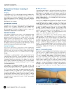
We prefer to use small-diameter interference screws with a double palmaris longus tendon graft for this procedure. Such screws are typically 15 mm in length and 4.75, 5.5, or 6.25 mm in diameter, depending on the diameter of the graft. One screw is inserted at the supinator crest, and another is inserted at the isometric point (Figs. 1 to 5). This kind of fixation is easy and fast and will allow for suitable strength and resistance for cycling load, similar to bone tunnels and superior to anchors. If the palmaris longus tendon is absent, regardless of the technique that is chosen, then the gracilis or semitendinosus tendon can be used. Allografts also can be used.
01 Harvesting the ipsilateral palmaris longus autograft.
38 ISAKOS NEWSLETTER 2019: VOLUME I
