
Page 37 - ISAKOS 2019 Newsletter Volume 1
P. 37
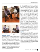
01 Tests used for hip microinstability.
It is important to keep in mind that hip microinstability may be associated with FAI and hip dysplasia. Hip instability may be associated with hip dysplasia, particularly if the Tönnis angle is high. Recently described radiographic signs have been used to identify hip instability. The Femoro-Epiphyseal Acetabular Roof (FEAR) index is defined by two lines. One is a line crossing the femoral head physeal scar, and the other is a line parallel to the sclerotic portion of the sourcil (similar to the Tönnis angle). A FEAR index of >5° is suggestive of instability. The Cliff sign, demonstrating a loss of femoral head offset, also has been seen in patients with hip instability. Magnetic resonance imaging (MRI) of the hip is used to check for labral tears and chondral status. In addition, magnetic resonance arthrography (MRA) can be performed to increase the sensitivity for labral tears. The MRA can include an injection of anesthetic, along with the dye, acting as a diagnostic test for an intra-articular cause of the pain. Typically, a decrease of 50% of the pain during the anterior impingement sign is considered a positive diagnostic test. Signs of instability on MRI or MRA include capsular defects, thin anterior hip capsule in the area lateral to the zona orbicularis on the axial view, and ligamentum teres tears.
Moreover, hip microinstability can appear as a complication after hip arthroscopy and is one of the main causes of postoperative pain. Microinstability occurs in cases in which the surgeon did not close the hip capsule and/or performed a large capsulotomy or even a capsulectomy, jeopardizing the function of the iliofemoral ligament. The clinical presentation of postoperative microinstability is similar that in nonoperative cases, with the patient complaining of anterior hip pain and presenting with similar findings on physical examination. An MRI may demonstrate a capsular defect in the anterior aspect of the hip.
Treatment
Initially, conservative treatment is instituted. The patient is advised to refrain from physical activities and to avoid hip rotation and deep flexion (i.e., squatting). A course of anti- inflammatory drugs may be prescribed to help alleviate acute pain. Physical therapy (PT) is the mainstay of this conservative trial. PT focuses on strengthening the hip girdle and stabilizing the hip joint. Core exercises are also important. It is necessary for the patient to be compliant with the proposed exercise regimen and to perform home exercises to complement the formal PT sessions. The idea is similar to that in cases of multidirectional atraumatic instability of the shoulder, in which a strong and developed musculature can overcome an unstable joint.
CURRENT CONCEPTS
  01A Abduction-hyperextension-external rotation test.
  01B Prone instability test.
Radiographic Evaluation
01C Hyperextension-external rotation test.
Radiographic evaluation includes an anteroposterior (AP) pelvic radiograph, a false-profile radiograph of the hip, and a profile radiograph of the proximal part of the femur. Many lateral views of the femur can be used, such as the Dunn view and the cross-table lateral view. On the AP pelvic radiograph, the clinician measures the joint space and observes any signs of subchondral sclerosis, which may indicate early degenerative disease. It is important to look for signs of FAI: the cross-over sign on the acetabular side is suggestive of pincer impingement, whereas loss of offset at the femoral head-neck junction is suggestive of cam impingement. The center-edge angle is used to measure the depth of the acetabular socket. Values of >40° demonstrate a global overcoverage typical of pincer impingement, and values of <25° demonstrate hip dysplasia (with values of 20° to 25° indicating borderline dysplasia).
 ISAKOS NEWSLETTER 2019: VOLUME I 35
