
Page 39 - ISAKOS 2018 Newsletter Volume 2
P. 39
CURRENT CONCEPTS
01
The first MRI or CT scan is performed without any stress applied in order to record the rest position. A second sequence is performed with posteroanterior stress applied to the proximal part of the calf by means of an ascending platform that is activated by a plunger under a 4-bar pressure. Then, the rotatory stresses (i.e., maximum internal rotation and maximum external rotation of the tibia) are tested independently by applying pressure through a rotatory footplate. Stress is gradually increased to comply with the viscoelastic mechanical characteristics of human tissues in order to ensure patient safety. Whenever possible, this procedure is performed bilaterally for side-to-side comparison.
Measurement Procedures
The MRI or CT scans are recorded with 1 to 5 mm slices. The laxity measurements (in mm) are made in both the medial and lateral tibiofemoral compartments. The slice selection is made independently for the medial and lateral tibial plateaus. The reference for the medial compartment is the first appearance of the medial portion of the proximal origin of the gastrocnemius muscle, and for the lateral compartment, the most medial sagittal aspect of the fibula. The measurements are made by calculating the distance between a line (oriented perpendicular to the tibial slope) crossing the most posterior point of the tibial plateau and a second line (oriented parallel to the first line) crossing the most posterior point of the femoral condyle4. Measurements are made with the knee at rest (non-stressed condition), as well as with the application of posteroanterior and rotatory stresses, for both the medial and lateral knee compartments. The difference between the rest and stressed conditions determines the amount of tibial displacement. Moreover, with the PKTD method, it is possible to calculate 2 combined measures in order to enhance the sensitivity and specificity of complete ACL ruptures. Global anterior translation (that is, the sum of the anterior tibial displacement of the medial and lateral tibial plateaus) provides high specificity (93%) with a cut-off of 15.1 mm. Global rotation (that is, the sum of the internal and external rotation of the lateral tibial plateau) has a high sensitivity (94%) with a cut-off of 11.1 mm3.
Clinical Relevance
The PKTD evaluation allows for the correlation of ligamentous “anatomy” and “function” within the same examination, which makes the PKTD a useful tool for the objective assessment of knee ligament injuries. The PKTD method can be used to assess differential diagnoses, to refine surgical indications, to assist with preoperative planning, to evaluate follow-up treatment outcomes, and to provide an additional criterion for return-to-sports decision-making. Additionally, it is possible to use this method to develop rotatory and sagittal laxity profiling of total ACL tears (Fig. 2), partial ACL tears (Fig. 3), combined ACL and posterolateral corner injuries (Fig. 4), and isolated posteromedial corner injuries (Fig. 5).
02
This method also can be used to demonstrate a “chewing- gum” effect (a situation in which a complete ACL rupture initially appears to be a partial rupture on MRI scans but, when stressed is applied by pushing the tibia anteriorly, a clear separation or an elongation of the ACL is seen). This effect may reveal that a ligament that would otherwise be considered intact and functional is actually non-functional.
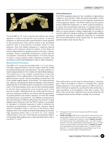
01 Photograph of the Porto Knee Testing Device (PKTD; Soplast, Valongo, Portugal).
02 Panels A through F: MRI scans showing PKTD measurements in the medial plateau (Panels A, B and C) and in the lateral plateau (Panels D, E and F) in a patient with a total ACL rupture. Panels A, B, and C: The measurements in the medial plateau were -1 mm with no stress (Panel A), 9 mm with posteroanterior stress (Panel B), and 13 mm with maximum external rotation of the tibia (Panel C). Panels D, E, and F: The measurements in the lateral plateau were 1 mm with no stress (Panel D), 11 mm with posteroanterior stress (Panel E), and 12 mm with maximum internal rotation of the tibia (Panel F).
ISAKOS NEWSLETTER 2018: VOLUME II 37
