
Page 31 - ISAKOS 2018 Newsletter Volume 2
P. 31
As severe (grade III-IV) untreated chondral disease is a contraindication to MAT, meniscal deficiency is also a contraindication to cartilage restoration. Similarly, the combined procedures enhance the outcomes of each other. The treatment choice for cartilage restoration is lesion-specific and typically is performed through an open approach after arthroscopic or arthroscopic-assisted MAT. Additionally, surgeons can complete the anterior horn MAT fixation during the open portion of the procedure (Fig. 2).
Graft Sizing
Preoperative MAT graft sizing is critical. Errors can result in improper biomechanics, meniscal extrusion, and failure. Grafts should be within 10% of the size of the native meniscus. If necessary, it is always preferable to use an undersized graft rather than an oversized one because of the increased risk of meniscal extrusion and early failure associated with oversized grafts. Additionally, proper mediolateral sizing is more important than anteroposterior sizing. For medial MAT, the Pollard x-ray method may be used for the determination of length and width. For lateral MAT, the Yoon equation for length and the arthropometric method for width may be preferred. If cost is not a concern, contralateral magnetic resonance imaging is accurate and useful for sizing.
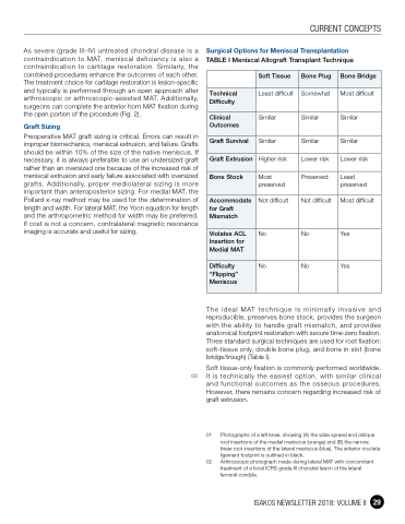
Surgical Options for Meniscal Transplantation
TABLE I Meniscal Allograft Transplant Technique
CURRENT CONCEPTS
Soft Tissue
Bone Plug
Bone Bridge
Technical Difficulty
Least difficult
Somewhat
Most difficult
Clinical Outcomes
Similar
Similar
Similar
Graft Survival
Similar
Similar
Similar
Graft Extrusion
Higher risk
Lower risk
Lower risk
Bone Stock
Most preserved
Preserved
Least preserved
Accommodate for Graft Mismatch
Not difficult
Not difficult
Most difficult
Violates ACL Insertion for Medial MAT
No
No
Yes
Difficulty “Flipping” Meniscus
No
No
Yes
02
The ideal MAT technique is minimally invasive and reproducible, preserves bone stock, provides the surgeon with the ability to handle graft mismatch, and provides anatomical footprint restoration with secure time-zero fixation. Three standard surgical techniques are used for root fixation: soft-tissue only, double bone plug, and bone in slot (bone bridge/trough) (Table I).
Soft tissue-only fixation is commonly performed worldwide. It is technically the easiest option, with similar clinical and functional outcomes as the osseous procedures. However, there remains concern regarding increased risk of graft extrusion.
01 Photographs of a left knee, showing (A) the wide spread and oblique root insertions of the medial meniscus (orange) and (B) the narrow, linear root insertions of the lateral meniscus (blue). The anterior cruciate ligament footprint is outlined in black.
02 Arthroscopic photograph made during lateral MAT with concomitant treatment of a focal ICRS grade-III chondral lesion of the lateral femoral condyle.
ISAKOS NEWSLETTER 2018: VOLUME II 29
