
Page 35 - ISAKOS 2018 Winter Newsletter
P. 35
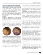
Chronic Exertional Compartment Syndrome
Lower extremity differential diagnosis: medial tibial stress syndrome (MTSS) (shin splints) and tibial stress fractures are the most common etiologies to differentiate from CECS in lower extremity. The differential diagnosis should also include evaluations for nerve entrapment (SPN, DPN, Sural N), deep vein thrombosis, popliteal artery entrapment and rarely some other clinical entities affecting bone or soft tissues.
Upper extremity differential diagnosis: a broad differential diagnoses includes peripheral nerve entrapment, peripheral neuropathy, motor neuron pathologies, and muscular disorders.
Treatment
Conservative: the reported results of non-operative management are modest and conservative interventions have generally been unsuccessful in restoring active patients to pain-free physical activity.
Surgery: when conservative measures are ineffective, or patients are unwilling to accept permanent activity or duty restrictions, operative management may be considered, with decompression of all symptomatic compartments. Fasciotomy is the elective treatment of CECS although selective use of partial fasciectomy for primary or revision cases may also be considered.
During the posterior compartment release, the saphenous vein and nerve must be identified and protected; the superficial fascia is easily identified, whilst the deep posterior compartment must be incised through the periosteum of the posterior medial border of the tibia. Hemostasis should be monitored carefully without tourniquet at the end of the procedure.
In the forearm, unlike the fascial compartments in the leg, pressure transfer from one compartment to the other appears to be substantial, thus, although the most affected muscles are probably the flexor digitorum profundus and flexor pollicis longus (deep volar compartment), superficial fasciotomy is adequate to release the entire forearm using a 4 to 8 cm longitudinal incision over the flexor compartment in the mid-forearm; if preferred, blunt splitting of the flexor digitorum muscle can be performed to allow access to the deep flexor muscles surrounding the ulna.
The affected compartment dilemma: in the lower leg, CECS is bilateral in 75–90% of the patients therefore, isolated release of one extremity may leads to subsequent contralateral surgery. The anterior compartment is most frequently affected (42%–60%) followed by the lateral (35%–36%), deep posterior (19%–60%), and the superficial posterior (3%–21%). Ideally, only the affected compartments should be released but there is a risk of failure and recurrence as untreated compartments occasionally becomes symptomatic once the most affected ones were released.
Complications
Perioperative complications often manifest early as local surgical site infections, wound healing difficulties, hematoma or seroma development and / or iatrogenic neurovascular injury, most commonly, the SPN and saphenous nerve.
Recurrences oscillates around 15 – 20% and typically occur after an initial symptom-free interval; it may result from inadequate release, failure to decompress a symptomatic compartment, postsurgical fibrosis or nerve compression. Entrapment of the SPN has been found in 44% of subjects presenting with recurrent CECS. Deep posterior compartment release success is lower (around 60%); the “fifth compartment” consisting of the tibialis posterior muscle which has its own fascia, when left intact can contribute to the inferior outcome observed in patients with deep posterior CECS.
Athletes performance
Postoperatively, activities are permitted as tolerated, expecting complete recovery in between 8 to 12 weeks. The best outcome is reported 14 months postop on average, most commonly in patients undergoing isolated anterior and lateral release. Recurrence rate is slightly higher for young patients, around 20%, while in adults is close to 11%.
CURRENT CONCEPTS
   As popularized by Mubarak and Owen, four-compartment fasciotomy of the leg can be performed through a dual- incision technique; the medial incision lies 2 cm medial to the tibia and the lateral incision is midway between the tibial crest and fibula. In recent years, several techniques for endoscopic fasciotomy, using smaller distal and proximal incisions, have also been described with encouraging results; specific long retractors, scissors and extended endoscope, as the ones used in vascular / general surgery are especially useful in this case.
When releasing the anterior / lateral compartments distally care must be taken to avoid injury of the superficial peroneal nerve (SPN) that exists the crural fascia at this level.
 ISAKOS NEWSLETTER 2018: VOLUME I 33
