
Page 26 - ISAKOS 2020 Newsletter Volume 2
P. 26
CURRENT CONCEPTS
Current Concepts in Managing Ankle Syndesmosis Injuries
Introduction and Popularity of Flexible Fixation Techniques
Over time, flexible fixation (e.g., suture-button) devices and hybrid constructs have been introduced and tested as an option to stabilize syndesmotic injuries. Small Level- IV studies showed good results with suture buttons for syndesmosis fixation, with no evidence of broken implants, earlier return to work, and a decreased need for hardware removal. In 2010, Bava et al. reported that flexible fixation was used in 10% of procedures. Subsequent Level-I and II studies comparing flexible fixation techniques and rigid screws demonstrated a number of common themes— namely, that suture buttons were associated with the same or better clinical outcomes compared with screws, that suture buttons were associated with a much lower rate of malreduction compared with screws, and that the adequacy of reduction was associated with better outcomes.
Shimozono et al., in a meta-analysis of randomized controlled trials evaluating the outcomes of screw versus suture-button fixation, found that the suture-button technique was associated with significantly improved functional outcomes, lower rates of broken implants, reduced need for removal of hardware, and fewer incidents of joint malreduction compared with the surgical screw technique.4 All of these constructs successfully repair the syndesmotic gap but fail to restore native joint motion3 or to address the rotational changes of the talus (Hunt et al., unpublished data). The clinical implications of these findings have yet to be determined; however, they suggest that a greater understanding of the syndesmosis during dynamic movements is necessary.
Restoring Biomechanics of the Ankle Syndesmosis
It is important to understand that most of the available clinical outcome studies on syndesmotic injuries involve patients with fractures that are associated with disruption of the syndesmosis, typically supination-external rotation injuries. It is also important to realize that an elite athlete with an unstable ligamentous injury of the syndesmosis will benefit from restoration of normal syndesmotic mechanics. We performed, in our laboratory, an in vivo assessment of normal three-dimensional (3D) mechanics under normal athletic conditions. In that study, we found that the shifts of the fibula at the level of the syndesmosis were largely posterior and external-rotation shifts rather than simple lateral widening. In fact, very little lateral widening can occur in an intact, uninjured syndesmosis. Instead, normal function during athletic activity is conveyed through rotation, anteroposterior, and superoinferior position shifts. No syndesmosis construct has been shown to restore normal mechanics following an unstable syndesmosis injury.
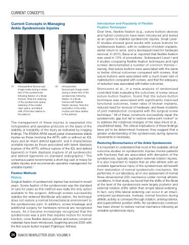
01 Intraoperative fluoroscopic image made during a stress test of the syndesmosis following fixation of a fibular fracture. Note the widening of the syndesmosis space, widening of the medial clear space, and lateral shift of the talus relative to the plafond.
02 Intraoperative fluoroscopic image made
during a stress test of the syndesmosis following fixation of a fibular fracture with flexible fixation devices. Note the restoration of the ankle mortice and talus relative to the plafond.
The management of these injuries is separated into nonoperative and operative protocols on the basis of the stability or instability of the injury as indicated by imaging findings. The ESSKA-AFAS expert panel characterizes stable injuries as those involving the AITFL with or without an IOL injury and an intact deltoid ligament, and it characterizes unstable injuries as those associated with latent diastasis (rupture of the AITFL without rupture of the IOL and deltoid ligament) or frank diastasis (rupture of all syndesmotic and deltoid ligaments on standard radiographs).2 The consensus panel recommends a short-leg cast or brace for stable injuries and recommends operative management for unstable injuries.2
Fixation Methods
History
Surgical fixation of syndesmotic injuries has evolved in recent years. Screw fixation of the syndesmosis was the standard of care for years as this method was really the only option available to the surgeon. Although screws successfully stabilize the widened tibiofibular articulation, the technique does not restore a normal biomechanical environment to the syndesmosis joint. In addition, screw breakage and additional surgery for hardware removal are recognized issues. As it became increasingly recognized that the syndesmosis was a joint that requires motion for normal function, more flexible device options and suture construct materials have been introduced, beginning around 2005 with the first suture-button implant (Tightrope; Arthrex).
24 ISAKOS NEWSLETTER 2020: VOLUME II
