
Page 19 - 2020 ISAKOS Newsletter Volume I
P. 19
CURRENT CONCEPTS
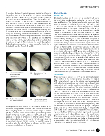
A specially designed measuring device is used to determine the defect size, and the scaffold is trimmed accordingly to fill the defect. A probe can be used to manipulate the implant into the correct position. When the scaffold is in place, it is sutured to the remnant of the host meniscus with an all-inside or inside-out technique. We prefer an all- inside suture technique because it is faster and avoids the need for additional approaches to retrieve sutures. In particular, all-inside vertical stitches are placed every 4 to 5 mm to suture the scaffold to the host meniscal remnant along the periphery, and horizontal stitches are used to fix the scaffold to the anterior and posterior border. The anterior and posterior extremities of the scaffold are fixed to the native remnant with horizontal stitches, and vertical stitches are placed every 5 to 10 mm along the scaffold periphery. Following the completion of suturing, scaffold stability is tested with a probe (Figs. 1, 2, and 3).
Clinical Results
Medial CMI
Clinical studies on the use of a medial CMI have demonstrated good results, particularly in terms of knee pain and function. The first series of medial meniscus CMI implants was described in the literature in 1997 by Stone et al., who reported no adverse effects and improved clinical scores at the time of the 3-year follow-up. Zaffagnini et al., in a prospective, non-randomized study, showed that a medial CMI provided better subjective outcomes scores and a lower VAS pain score in comparison with the findings in a group of matched controls who were managed with partial medial meniscectomy. Rodkey et al., in a multicenter prospective clinical trial of 311 patients who were managed with either a medial CMI or partial meniscectomy, reported that no significant clinical differences were observed between the groups at the time of the 5-year follow up in the “acute” arm of the study (comprising patients with no previous meniscal procedures) whereas the CMI group demonstrated some improvement in the Tegner index in the “chronic” arm of the study (comprising patients with a history 1 to 3 meniscal procedures). Monllau et al., in a study of 22 patients who were followed for a minimum 10 years after treatment with the CMI, reported significant pain relief and functional improvement. In other studies, medial CMI implantation has been associated with good clinical results in terms of knee function and pain reduction after follow-up periods ranging from 6 months to 10 years, particularly when used for the treatment of chronic partial lesions of the medial meniscus.
Lateral CMI
The accumulated experience with lateral CMI implantation as described in the literature is less than that for medial CMI implantation. However, the clinical results associated with lateral meniscectomy have been worse than those associated with medial meniscectomy because of the challenging biomechanics of the lateral meniscus, including a small contact area, high peak stresses, and extreme mobility. Therefore, a meniscal preservation procedure should be attempted whenever possible, and, in cases of partial meniscal loss, a CMI scaffold represents a viable alternative. Hirschmann et al., in a series of 67 patients, showed significant improvements in terms of subjective and objective outcomes after both lateral CMI implantation (12 patients) and medial CMI implantation (55 patients), with no difference in outcomes between the groups. Zaffagnini et al., in a prospective multicenter study of 43 patients with partial meniscal defects or irreparable meniscal tears, reported that lateral CMI implantation significantly improved knee function and reduced knee pain in the short term. Therefore, despite shorter experience with lateral CMI implantation, the clinical results are similar to those reported in association with the medial scaffold, with satisfactory clinical improvement at 2 years of follow up.
01 Loss of meniscal tissue in previous lateral
meniscectomy.
02 Quantification of the defect size.
03 Fixation of the scaffold with-all inside stitches.
04 Fixation of the scaffold with-all inside stitches.
In the postoperative period, a knee brace, locked in full extension, is applied for 1 month. The patient removes the brace only to perform self-assisted passive range-of-motion (ROM) exercises. ROM is limited to a gradual range of 0° to 90° in the first month. Unlimited ROM, with active and passive exercises, is encouraged after 5 weeks. Weight- bearing is not allowed for 3 weeks. Partial weight-bearing is permitted between 4 and 5 weeks. Full weight-bearing is permitted after 5 weeks. Return to full sport activities is not recommended before 4 to 6 months.
ISAKOS NEWSLETTER 2020: VOLUME I 17
